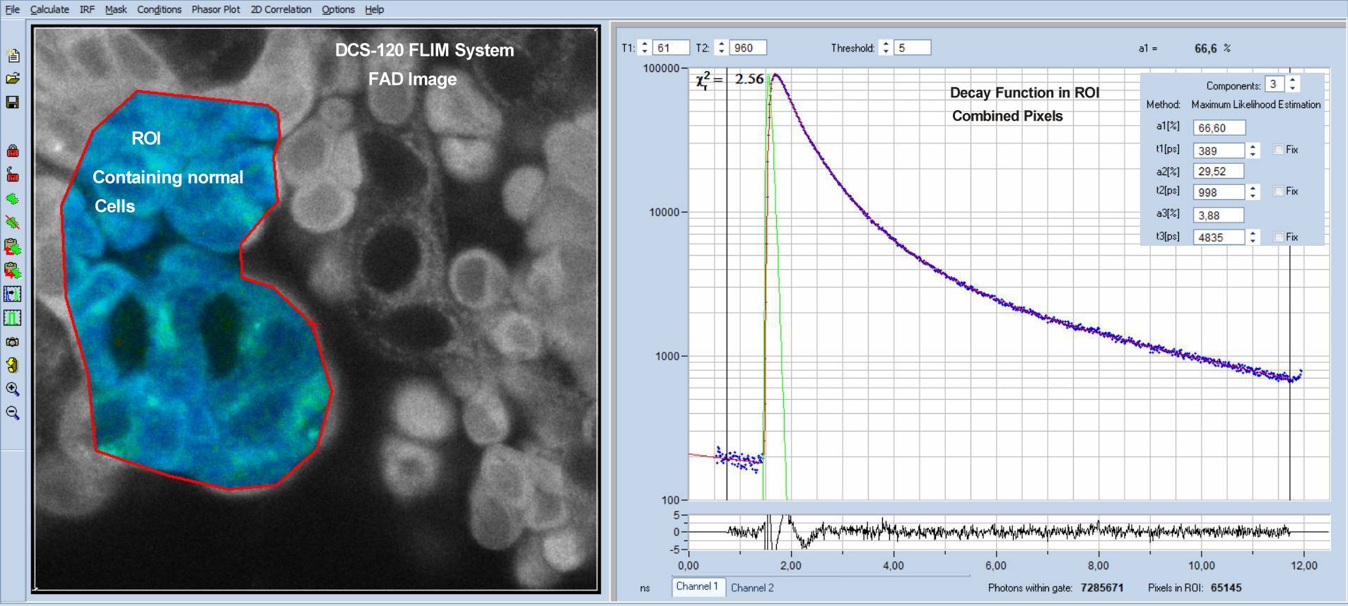
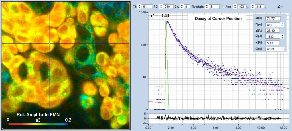
In contrast to NAD(P)H data, metabolic-FLIM data derived from FAD images are often inconclusive. It has long been suspected that FAD data are biased by a fluorescence decay component from FMN. We therefore checked for possible traces of FMN fluorescence in FLIM data recorded by our DCS-120 metabolic FLIM system.
Indeed, we found a decay component with a lifetime around of 4.3 ns to 5.5 ns which cannot be attributed to FAD but very well to FMN. The amplitude of the FMN component was 3.9% to 5% in normal cells and 8.8% to 10% in cancer cells. We are finding the FMN component both in data from larger ROIs with high photon numbers and in pixel data with large binning factors. The presence of a decay component from FMN may have implications to the use of FAD data for cancer diagnosis, especially if the the amplitude ratio, a1/a2 or the FLIRR ratio is used as cancer indicators. Certainly, an amplitude contribution of 3.8% or even 8.84% does not render the a1/a2 ratio or the FLIRR ratio useless. It can, however, introduce unpredictable shifts in these parameters.
Please see Application Note for more information.