Table of Contents:
- Applications of Fluorescence Lifetime Imaging Microscopy
- Molecular Imaging
- Metabolic Imaging
- FRET Imaging
- Simultaneous NAD(P)H, FAD and pO2 Imaging
- FLIM-Based In Vivo Imaging
- In-Vivo Diagnostics of Skin
- Fluorescence-Lifetime Ophthalmoscopy (FLIO)
- Personalized Chemotherapy
- Leading Technology for All Your FLIM Needs
Applications of Fluorescence Lifetime Imaging Microscopy
From the beginning, bh Fluorescence Lifetime Imaging Microscopy (FLIM) devices have been designed with application in Life Sciences in mind. High sensitivity, high photon efficiency, high time resolution, fast recording, and the ability to resolve multi-exponential decay functions are the basic requirements in these applications. Moreover, bh systems address the complexity of live systems by their ability to record several biological parameters simultanously and in their mutual depencence. With functions like simultaneous FLIM and PLIM, parameter-controlled mosaic FLIM, multi-wavelength FLIM, or triggered recording of fast physiological effects bh’s multi-dimensional TCSPC technique addresses these needs almost perfectly. These features make bh FLIM systems superiour to other systems on the market.
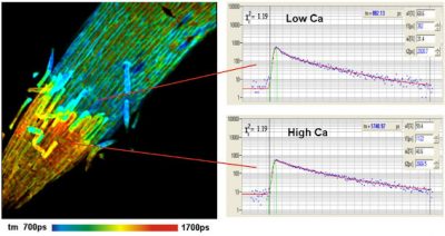
 The basis of all these applications is the fact that the fluorescence lifetime of most fluorophores is depending on their molecular environment. Accurate measurement of the fluorescence lifetimes or, more exactly, the fluorescence decay functions in the pixels of a FLIM image is therefore used to obtain reliable information on biological systems. An inherent advantage of FLIM in this respect is that the fluorescence lifetime, in contrast to intensity, does not depend of the concentration of the fluorophore, the laser power, the detector gain, or other experimental or instrumental details. An example of a Calcium-concentration measurement is shown in the figure on the right.
Although the bh FLIM technique is perfectly suited for entry-level applications it develops its full power in advanced applications such as:
Molecular Imaging
‘Molecular Imaging’ includes a wide range of applications, such as pH measurement in tissues, cells, and subcellular compartments, concentration of physiologically relevant ions, such as Ca++, Mg++, Na+, K+, Cl–, or Oxygen concentration, or Glucose concentration.
Metabolic Imaging
Metabolic Imaging uses the decay functions of NAD(P)H and/or FAD to determine the metabolic state of cells and tissues. Here, the information is not primarily in the fluorescence lifetimes but in the multi-exponential composition of the decay.
FRET Imaging
FRET (Förster Resonance Energy Transfer) measurements use the energy transfer from a donor to an acceptor molecule to probe protein conformation and interaction between different proteins. Also here, the fluoresecnce decay functions are multi-exponential, and the FLIM technique has to resolve the parameters of the complex decay.
Simultaneous NAD(P)H, FAD and pO2 Imaging
The bh technique can combine metabolic imaging with simultaneous pO2 imaging. With this combination, the metabolic state of a cell can be investigated in dependence of the oxygen concentration.
FLIM-Based In Vivo Imaging
The methods of Fluorescence Lifetime Imaging Microscopy (FLIM) are particularly suited for in-vivo diagnostics as they are noninvasive and non-destructive. Thus, they are widely used for clinical research and medical examination on live subjects. Examples are given below.
In-Vivo Diagnostics of Skin
Multiphoton-lifetime tomography of human skin uses two-photons excitation in combination with non-descanned detection. Multi-photon FLIM delivers optically sectioned images of tissue layers as deep as 100 µm. In-vivo two-photon imaging of human skin cells is possible without impairing the viability of the tissue. From z-stacks of FLIM images three-dimensional structures can be reconstructed at sub-cellular resolution.
Fluorescence-Lifetime Ophthalmoscopy (FLIO)

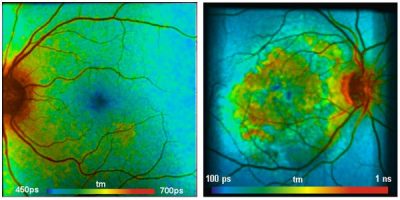
Ophthalmic FLIM uses a combination of fast beam scanning and excitation by a picosecond diode laser. This method is so sensitive that it is able to record lifetime images of the fundus (background) of the human eye. By this, the early discovery of eye diseases is possible, as these are often accompanied by metabolic changes of the fundus. In turn, these cause changes in the fluorescence decay parameters of endogenous fluorophores.
Personalized Chemotherapy
To target the special type of cancer a patient is suffering from, it is crucial to find the most efficient anti-cancer drug. The response of cancer cells to the different types of drugs is not entirely predictable though. Therefore a biopsy is taken, the cells are cultured and treated with different drugs. At the same time they are repeatedly imaged by FLIM. Fluorescence lifetimes indicate early shifts in the metabolic state of a cell after the treatment. Thus, with these measurements the most efficient medication can be determined within only a few days.
Leading Technology for All Your FLIM Needs
Choose the ideal equipment for your laboratory or clinic from our complete high-grade FLIM systems or our versatile upgrades for your existing microscope and scanner. You need further information on which solution would be best for your desired application? Send us an inquiry via our contact form or call us at +49 (30) 212 80 02-0!
FLIM Upgrade Kits for Laser Scanning Microscopes

As a technology leader in TCSPC and TCSPC FLIM since 1993, Becker & Hickl offer a wide range of high-performance Fluorescence Lifetime Imaging (FLIM) systems for laser scanning microscopes. Our systems have found broad application in Molecular Imaging in Life Sciences, Metabolic FLIM, Clinical FLIM, Diffuse Optical Tomography (DOT), Fluorescence Correlation (FCS, FCCS), Förster Resonance Energy Transfer (FRET) and more. Using our proprietary multidimensional time correlated single photon counting (TCSPC) and TCSPC FLIM techniques our systems are characterized by ultra-high time-resolution, near-ideal photon efficiency, and the capability of ultra-fast Time-Series FLIM, Mosaic FLIM, Multi-Wavelength FLIM, Excitation-wavelength multiplexed FLIM, and Simultaneous FLIM and PLIM. Choose your FLIM Upgrade now:

