The bh DCS-120 MACRO detects tumors in mice via FLIM of endogenous NAD(P)H. NAD(P)H exists in a bound and a free form. The free-to-bound ratio changes with the metabolic state. Since bound and free NAD(P)H have different fluorescence lifetimes a shift in the metabolic state results in a change in the decay profile. A shift to glycolysis yields a larger amplitude of the fast decay component and a shorter mean lifetime, a shift to oxydative a smaller amplitude of the fast component and a longer mean lifetime. To detect NAD(P)H images, the imaging system uses confocal scanning in combination with ps diode laser excitation and bh’s multi-dimensional TCSPC process. The object is placed directly in the image plane of the scanner. The system has inputs for two lasers, and two fully parallel detection channels. For NAD(P)H FLIM, a 375 nm picosecond diode laser is used for excitation. The emission is detected through a 440 to 475 nm bandpass filter. The system can be extended for simultaneous detection of FAD. In that case, a 405 nm laser is used in the second laser channel, and multiplexed with the 375 nm laser. FAD data are recorded in the second detection channel.
For more information please see:
- V. I. Shcheslavskiy, M. V. Shirmanova, V. V. Dudenkova, K. A. Lukyanov, A. I. Gavrina, A. V. Shumilova, E. Zagaynova, W. Becker, Fluorescence time-resolved macroimaging. Opt. Lett. 43, No. 13, 3152-5155 (2018)
- Becker & Hickl GmbH, DCS-120 confocal and multiphoton FLIM systems, user handbook, 7th edition (2017)
- W. Becker, The bh TCSPC Handbook, 7th edition (2017)
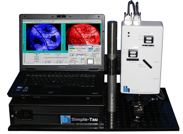
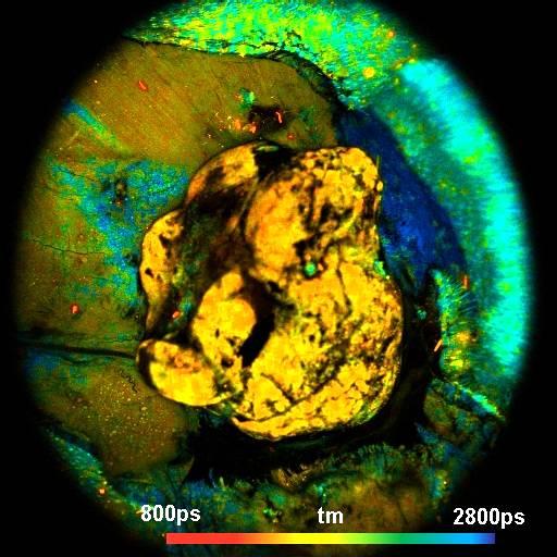
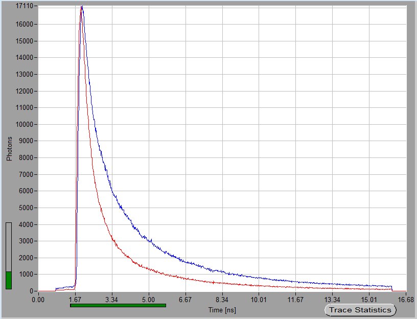
Figures, left to right: DCS-120 MACRO FLIM system. Tumor in a mouse, size of image 15 x 15 mm. Fluorescence decay curves from area within tumor (red) from area within normal tissue (blue).