
Overview
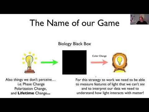
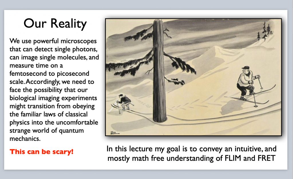
Förster resonance energy transfer (FRET) is a widespread microscopy method used for measuring molecular proximity on a 1-10 nm range. I attribute its popularity to several factors, but primarily to the development of genetically encoded biosensors, and the ability to genetically tag proteins with fluorescent proteins and study their interactions inside living cells and organisms. While many microscopy techniques have been developed to measure FRET, fluorescence lifetime microscopy (FLIM) is in many ways the simplest, and most straightforward approach. Nonetheless, both FRET and FLIM can be intimidating techniques to master, particularly for new practitioners, as they involve monitoring kinetic events that typically occur in the picosecond to nanosecond (or faster) range and are used to probe events involving the interactions of single photons with one or more fluorophores. These types of events occur at the interface of classical physics and quantum mechanics. In this webinar I will present a primer on FRET and FLIM basics using minimal math and hopefully generating maximal intuition. My goal is to help new practitioners feel comfortable thinking about the relevant events occurring on a nanoscale and in so doing motivate and empower biologists to consider FLIM-FRET for the powerful research tool it can be.
About the Speaker
Dr. Vogel received his B.S. from the City College of New York in 1978 and his Ph.D. in Biochemistry and Molecular Biophysics from Columbia University in 1989, where he studied G-proteins in the nervous systems of Aplysia and squid with Dr. James H. Schwartz. During a postdoctoral fellowship at NIH, he investigated the calcium dependence of exocytosis in sea urchin eggs using light microscopy and discovered heterogeneity for calcium responses in a population of secretory vesicles. Dr. Vogel next became an assistant and subsequently a tenured associate professor at the Medical College of Georgia (1997-2003). Using two-photon microscopy he studied the mechanism of exocytosis – endocytosis coupling, as well as the role plasma-membrane wound repair plays in muscular dystrophy. He joined NIAAA/NIH as an investigator in 2003 and became a senior investigator in 2013. His laboratory develops new fiber optic and microscope-based assays using FRET, fluorescence polarization, and fluorescence correlation spectroscopy to study protein-protein interactions in living cells, focusing on the structure and function of CaMKII holoenzyme. More recently Dr. Vogel’s lab has started investigating biological mechanisms relevant to quantum biology using photon antibunching, time resolved anisotropy, and circular dichroism. His lab uses fluorescent protein dimers as a model system for studying how excitonic coupling is possible in a hot and wet environment. Dr. Vogel is also a faculty member of the annual University of Virginia FRET Workshop.