
Overview
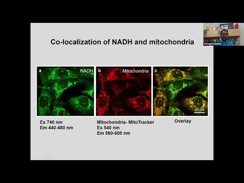
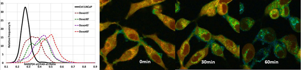
The seminal work by Chance in the 1960’s exploiting the auto-fluorescent properties of the coenzymes NADH and FAD for measuring the cellular reduction/oxidation (REDOX) states noninvasively since then the REDOX field has grown exponentially. The intensity-based methods were expanded followed by Fluorescence lifetime imaging (FLIM) assays to monitor REDOX changes as markers for changed metabolic states. These FLIM assays are of particular interest in measuring responses to treatment in various by oxidative phosphorylation (OXPHOS) and (often preferentially) by glycolysis. Two-photon fluorescence lifetime imaging microscopy (FLIM) is widely used to capture auto-fluorescence signals from cellular components to investigate dynamic physiological changes in live cells and tissues. Instead, the basic FLIRR (fluorescence lifetime redox ratio) assay is based entirely on lifetime measurements, unaffected by intensity artefacts, isolates individually segmented cell data, and separates intra-cellular OXPHOS and glycolysis data. In this talk, I will discuss the FLIRR approach for prostate cancer investigation in living cells.
About the speaker
Dr. Periasamy is the Director and founder of the internationally known WM Keck Center for Cellular Imaging. He is professor of Biology and Biomedical Engineering at the University of Virginia, Charlottesville, USA. A key area of Dr. Periasamy’s research is focused on the design and development of optical methodology including advanced light microscopy techniques to investigate/monitor exogenous and endogenous molecular interactions in live cells, tissues, and animals. Dr. Periasamy is one of the pioneers in the development of fluorescence lifetime imaging microscopy (FLIM) for measuring the oscillations in cytosolic and nuclear free calcium in single intact living cells. He developed a 2- and 3-color steady state, confocal, multiphoton, and FLIM based Förster resonance energy transfer (FRET) imaging systems for protein localization in living specimens. He has published 135 articles in refereed journals and book chapters. He has given 156 invited lectures nationally and internationally. Dr. Periasamy has edited three books, series book editor on cellular and clinical imaging (7 books), Chairperson in organizing an annual International conference on Multiphoton Microscopy in the Biomedical Sciences through SPIE (since 2001) and runs a hands on training annual workshop on FLIM & FRET Microscopy at the University of Virginia, Charlottesville during March (since 2002). Dr. Periasamy was elected “Fellow” members of the SPIE Optical Society in 2012.