
Overview
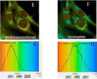
Multi-photon fluorescence lifetime microscopy (FLIM) is used as a versatile method to measure changes in mammalian metabolism by tracking the auto-fluorescent coenzymes NAD(P)H and FAD. In many human pathologies – including cancer – the metabolism is disrupted or reprogrammed. However, executing an optimized FLIM image acquisition process is my no means a trivial challenge, e.g. balancing acquisition time with laser power, avoiding potential photo-bleaching (and whether it actually affects lifetime parameters); can FLIM be applied to fixed specimens? These and other questions will be covered. The next step concerns a correct lifetime fitting process, followed by quantitative analysis which can be applied to complete ‘fields-of-view’, individually segmented cells to discover heterogeneous responses to treatment and tracking changes in oxidative phosphorylation based on NAD(P)H and FAD imaging. I am presenting practical details of the multi-step sequence of FLIM acquisition, choices made during fitting and analysis, based images generated by a time-correlated-single-photon-counting (TCSPC) system, fitted with Becker & Hickl software and further processed with open-source ImageJ/Fiji and Python software.
Speaker
After a 35 years business career in the pharmaceutical industry, Horst Wallrabe joined University of Virginia, Dep. of Biology in 1997 – at age 60 retirement – as a full-time Community Scholar. Developed expertise in molecular cell biology, FRET & FLIM microscopy resulting in >20 peer-reviewed papers, based on own and cooperative research. Engagement in undergraduate training and speaking engagements, representing the department are part of the daily activities.