Non-Descanned FLIM Detection
in Multiphoton Microscopes
Abstract. Multiphoton microscopes use a femtosecond NIR laser to excite
fluorescence in the sample. Excitation is performed via a multi-photon
absorption process. The advantage of multiphoton excitation is that
fluorescence is excited only in the focal plane, and that the laser light
penetrates deep into the sample. Multiphoton excitation becomes particularly
efficient in combination with non-descanned detection (NDD). This application
note describes the principles of non-descanned detection in combination with
the bh fluorescence lifetime imaging (FLIM) devices and shows a number of
typical NDD FLIM results.
Multiphoton Excitation
A multiphoton microscope excites the
fluorescence in the sample via simultaneous absorption of several (usually two)
photons of a femtosecond-pulsed near-infrared laser. Absorption and scattering
in the NIR is substantially lower than at visible or ultra-violet wavelengths.
Therefore multiphoton excitation penetrates substantially deeper into a sample
than the visible or UV laser of a confocal microscope, see Fig. 1, left and
middle. Moreover, depth resolution is obtained via the nonlinearity of the
excitation process, see Fig. 1, middle. In other words, fluorescence is excited
only in the focus of the femtosecond laser.
Fig. 1: Left: One-photon excitation. The effective excitation power
decreases rapidly with increasing depth. Middle: Two-photon excitation. The NIR
laser penetrates deeply into the sample. Right: The fluorescence from a deep
focus is scattered on the way out of the sample. It leaves the back aperture of
the microscope lens in a wide cone.
Principle of Non-Descanned Detection
When multiphoton excitation is used to
reach deep sample layers scattering of the fluorescence photons becomes
important, see Fig. 1, right. The scattering coefficient at the fluorescence wavelength
is higher than at the excitation wavelength. Thus, a large amount of the
fluorescence photons is scattered on the way out of the sample. These photons
leave the back aperture in a diffuse cone of light. Because the scattered
photons originate from the focus of the femtosecond laser they bear useful
information. However, scattered photons cannot be collimated and thus not be
fed back through the beam path of the scan head of laser scanning microscope.
The situation is shown in Fig. 2, left. Even
if the pinhole is opened wide mainly ballistic photons would reach the confocal
detector, scattered photons would be lost. The problem of detecting scattered
photons is solved by non-descanned detection (NDD). NDD makes use of the fact
that fluorescence in a multiphoton microscope is only excited in the focus. It
is therefore not necessary to use a pinhole to reject out-of-focus
fluorescence.
The principle of NDD is shown in Fig. 2,
right. A second dichroic mirror diverts the fluorescence light immediately
behind the microscope lens, and a transfer lens projects these photons on the detector.
With a sufficiently large transfer lens and a reasonably large detector area,
all photons emerging from the back aperture of the microscope lens are
transferred to the detector.

Fig. 2: Left: Confocal detection. Fluorescence scattered in the sample
does not reach the detector. Right: Non-descanned detection. Both ballistic and
scattered photons are transferred to the detector.
Details of a non-descanned detection beam
path are shown in Fig. 3. The fluorescence leaving the back aperture of the
microscope lens, L1, is separated from the excitation light by a dichroic
mirror. The fluorescence is collected by a transfer lens, L3. This lens
projects a de-magnified image of the back aperture of the microscope lens on
the active area of the detector. L3 is normally placed directly behind a side
port of the microscope. It should be large enough to collect all light
transmitted through the internal beam path of the microscope, and have a focal
length short enough to obtain a sufficiently small image of L1 on the surface
of the detector. With typical values, such as d1 = 180 mm, d4 =
50 mm, and D3 = 30 to 40 mm, a de-magnification of a factor of
3.6 is obtained. That means, a detector diameter of 2.8 mm is sufficient to
detect the light emerging from a microscope lens back aperture of 10 mm
diameter.
Fig. 3:
Details of NDD beam path
It should be noted that the transfer lens,
L3, does not need to have perfect optical correction. It is only required that
it concentrates the fluorescence light into an area smaller than the active
area of the detector. Usually a simple plano-convex or bi-convex lens is
sufficient. In extreme cases an aspherical lens can be used.
The location of L1 in the beam path, i.e.
the distance d1, has no relation to the location of the upper image plane. L1
can be placed at any distance, either before or behind the upper image plane.
However, L1 should not be placed directly in the upper image plane to avoid
dust on the lens causing shadows in the images recorded.
In any case, it must be avoided that the
detector is placed in an image plane conjugate with the sample. This would
result in scanning the luminescent spot over the detector and thus imprint
internal structures of the detector on the images [1, 2]. An NDD setup can
easily be checked by placing a sample in the microscope and turning on the lamp
in the transmission path. The spot in the detector plane is then examined on a
sheet of paper. In a correctly designed beam path the spot should be an image
of the microscope lens, not an image of the sample.
Practical Examples of NDD FLIM Detectors
As shown in Fig. 3, non descanned detection
requires a dichroic beamsplitter in the microscope main body, and a suitable
port to which the fluorescence light is directed. Suitable beamsplitters
positions and side ports are available for most research-grade microscopes. A
common problem of NDD FLIM is that light from microscope lamps can be
transmitted directly to a detector attached to one of these ports. The light
can be so strong that it can damage the detector. The non-descanned FLIM
detectors of bh are therefore equipped with overload shutdown, and electronically
controlled shutters [2, 7, 8] are available. The shutter assembly
also contains the lens that transfers the photons to the detector.
A bh detector/shutter assembly with a PMC‑100
cooled PMT module coupled to the side port of the IX81 microscope of an
FV 300 multiphoton system is shown in Fig. 4. Please see [11] for details.
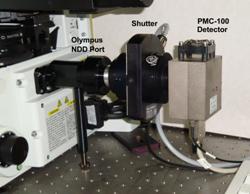
Fig. 4: PMC‑100 detector with shutter coupled to the right side port
of the IX81 microscope of an FV 300 multiphoton scanning system
bh NDD FLIM systems are available for
different microscopes and with a number of different detectors [8, 10, 11].
Examples are shown in Fig. 5. The bh PMC‑100-01 module with a Leica RLD
adapter is shown in Fig. 5, left. The PMC‑100 is a compact PMT module
containing the preamplifier, the PMT power supply, overload shutdown circuitry,
and thermoelectric cooling [2]. For ultra-high time resolution the Hamamatsu
R3809U MCP PMT is available. To provide maximum safety against overload the
R3809U is usually operated via a shutter, see Fig. 5, second left and middle.
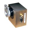
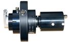
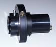
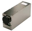
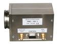
Fig. 5: Left to right: bh PMC‑100 detector module with Leica RLD
adapter, R3809U MCP PMT with shutter and Leica RLD adapter, R3809U MCP PMT with
shutter and Zeiss LSM 510 adapter, HPM‑100-40 hybrid detector module
with Zeiss LSM 710 adapter, PMZ-100 PMT module with Zeiss LSM 710
adapter.
Since 2009 bh have added ultra-sensitive GaAsP
hybrid detectors to their TCSPC systems. These detectors combine the
sensitivity of a single-photon avalanche photodiode with the large active area
of a PMT. They are free of afterpulsing and thus deliver an extremely high
dynamic range of fluorescence decay detection [5, 8]. A HPM‑100-40 with a Zeiss
LSM 710 NDD adapter is shown second right. A PMZ‑100 PMT module with
an LSM 710 NDD adapter is shown right.
Multi-spectral multiphoton NDD FLIM [6] is
available by using the bh MW FLIM multi-wavelength detector assembly [2, 4]. In combination with the bh TCSPC
technique, the MW FLIM detects simultaneously in 16 wavelength channels,
without any wavelength scanning or time gating [1, 2]. Multi-wavelength detection
requires the fluorescence light to be transferred into the entrance slit of a polychromator.
The bh multi-spectral FLIM assembly therefore uses a fibre bundle to achieve a
transformation of the cross section of the light bundle [1, 2, 4, 6]. The MW FLIM assembly attached to
the side port of an IX81 FV300 system [11] is shown in Fig. 6 .
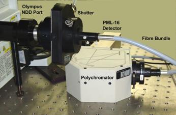
Fig. 6:
Multi-spectral FLIM assembly attached to the side port of an IX81 FV300
scanning system
FLIM Electronics
bh NDD FLIM systems for multiphoton
microscopes use the bh Simple-Tau 150 and Simple-Tau 152 TCSPC
systems. The systems contain one or two bh SPC‑150 TCSPC FLIM modules and
a bh DCC‑100 detector controller. The TCSPC electronics is contained in
an extension box that is connected to a lap-top computer via a bus extension
interface. Please see [1, 2] for details of the bh TCSPC technique. The Simple
Tau 150 and 152 Systems are shown in Fig. 7.
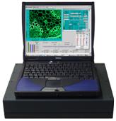

Fig. 7: Simple-Tau 150 and Simple-Tau 152 systems. One or two
SPC‑150 TCSPC-FLIM modules and a DCC‑100 detector controller are
contained in a bus extension box of a lap-top computer
The FLIM systems come with the TCSPC
hardware and the software readily installed. The software allows you to select
between a large number of image formats with different numbers of pixels and
time channels. Single FLIM images and time-series of FLIM images can be
recorded. For focusing and sample positioning, a fast preview mode allows you
to display fluorescence images of the sample as fast as 2 frames per second [8,
10, 9]. You can easily change between these setups by clicking on a button of
the Predefined Setup panel.
Data analysis includes fitting with single,
double and triple-exponential models. For samples with SHG components a
Scatter component can be included in the fit. The results are displayed by
false-colour images. Any parameter of the fit model, ratios of parameters, mean
lifetimes, average lifetimes, SHG components, and the FRET efficiency of the
interacting donor component of FRET experiments can be displayed. Please see [8] or [9] for details.
Typical Results
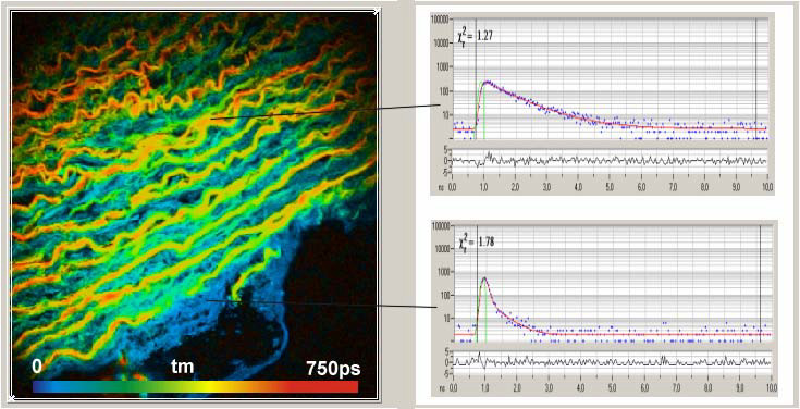
A typical FLIM result obtained with a
single-detector NDD FLIM system connected to an Olympus FV 300 multiphoton
microscope is shown in Fig. 8. The samples delivers both fluorescence from endogenous
fluorophores and an SHG signal from the collagen in the tissue. SHG is an
ultra-fast process; the collagen thus shows up via an infinitely fast signal
component.

Fig. 8: Heart tissue sample. The sample shows fluorescence from endogenous
fluorophores and SHG from the collagen in the tissue. Mean lifetime of double-exponential
decay, colour scale from 0 to 750 ps.
Fig. 9 shows NDD FLIM images recorded by a
dual-channel Simple‑Tau 152 FLIM system attached to a Zeiss
LSM 710 NLO. Two images were recorded simultaneously in two wavelength
intervals. The left two images show a pig skin sample excited at 800 nm,
and recorded at 400 to 480 nm (left) and 480 to 650 nm (right).
The left image shows the fraction of SHG, the right image the fluorescence
lifetime. The right two images show a plant sample. The images were recorded at
450 to 550 nm and 550 to 700 nm. The left image shows the flavines in
the cell membranes, the right image the chlorophyll in the chloroplasts of a
plant sample.
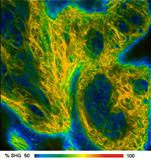
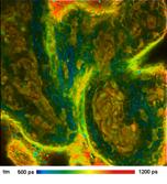
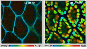
Fig. 9: Multiphoton images, LSM 710 NLO, dual-channel FLIM, HPM‑100‑40
GaAsP hybrid detectors. Left: Pig skin, 40 um deep, excitation
800 nm. Left image shows percentage of SHG at emission wavelength
<480 nm, right image shows fluorescence lifetime at >480 nm.
Right: Plant sample, excitation at 860 nm. Left image emission at 450 to
550 nm, right image emission at 550 to 700 nm.
A multi-wavelength FLIM result is shown in Fig.
10. The images show a plant sample in 16 wavelength intervals. The data were
recorded by a bh Simple-Tau 510 with bh multi-wavelength FLIM assembly
connected to the non-descanned output of a Zeiss LSM 710 NLO.
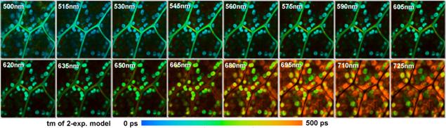
Fig. 10:
Multiphoton Multispectral NDD FLIM. LSM 710 NLO, bh MW FLIM detector
Summary
Multiphoton excitation penetrates
substantially deeper into a sample than one-photon excitation. The depth
penetration capability of multiphoton excitation can only be exploited by
non-descanned detection. Non-descanned detection systems project an image of
the back aperture of the microscope lens on the detector. With appropriate lens
diameters, NDD systems are not only highly efficient for ballistic photons,
they also detect photons scattered on the way out of the sample. Non-descanned
detectors for the bh FLIM systems are available for all commercial laser
scanning microscopes, and can easily be adapted to home-built systems.
References
1.
W. Becker, Advanced time-correlated single-photon counting techniques. Springer, Berlin,
Heidelberg, New York, 2005
2. W. Becker, The bh TCSPC handbook. 3rd edition, Becker
& Hickl GmbH (2008), available on www.becker-hickl.com
3. W. Becker, A. Bergmann, M.A. Hink, K. König, K. Benndorf, C. Biskup,
Fluorescence lifetime imaging by time-correlated single photon counting, Micr.
Res. Techn. 63, 58-66 (2004)
4.
Becker & Hickl GmbH, PML-16-C 16
channel detector head for time-correlated single photon counting. User
handbook. Available on www.becker‑hickl.com
5.
Becker & Hickl GmbH, The HPM‑100-40
hybrid detector. Application note, available on www.becker-hickl.com
6. W. Becker, A. Bergmann, C. Biskup, Multi-Spectral Fluorescence
Lifetime Imaging by TCSPC. Micr. Res. Tech. 70,
403-409 (2007)
7.
Becker & Hickl GmbH, DCC-100 detector
control module, manual, www.becker‑hickl.com
8.
Becker & Hickl GmbH, Modular FLIM
systems for Zeiss LSM 510 and LSM 710 laser scanning microscopes.
User handbook. Available on www.becker-hickl.com
9.
Becker & Hickl GmbH, DCS-120 Confocal
Scanning FLIM Systems, user handbook. www.becker-hickl.com
10.
bh NDD FLIM systems for Leica SP2 MP and
SP5 MP Multiphoton Microscopes. Application note, Becker & Hickl GmbH,
www.becker-hickl.com
11.
Non-Descanned FLIM Systems for Olympus FV-1000
and FV-300 Multiphoton Microscopes. Application note, Becker & Hickl GmbH
(2007), www.becker-hickl.com