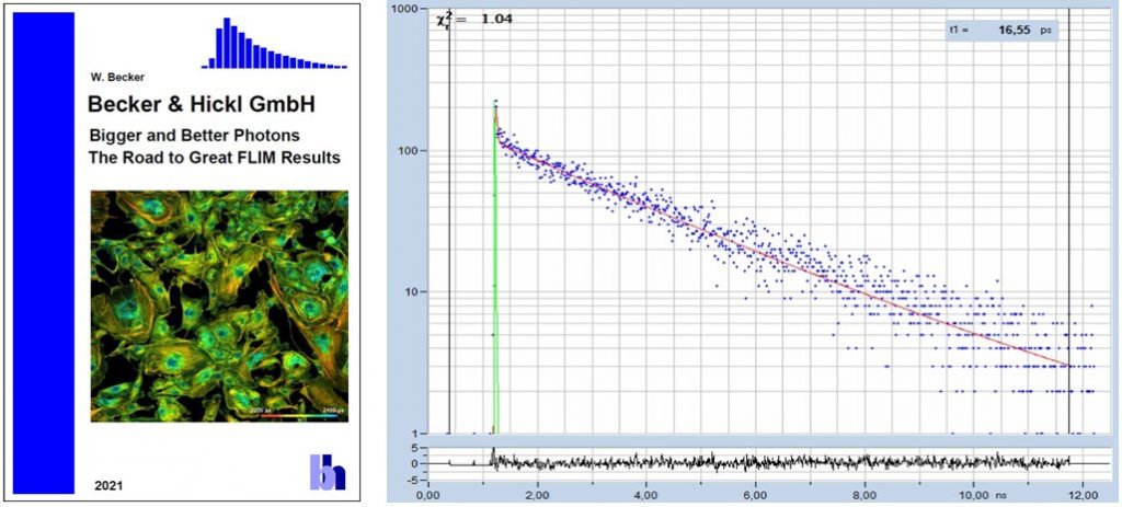
What makes a good FLIM image? It should have perfect spatial resolution, it should be recorded into a sufficiently high number of pixels, it should have high contrast, low background, no out-of-focus blur, and it should display the fluorescence lifetime at a high signal-to-noise ratio. Just as the images shown below. An experienced FLIM user will probably add that just recording the fluorescence lifetime is not enough, and the entire decay function should be recorded in every pixel.
Why do FLIM images published in scientific papers rarely look like the images shown below? There is actually no reason for that. All that has to be done is to use perfectly aligned optics, the right microscope lens, perfect focusing, the right excitation and detection wavelengths, the right detector, and a little bit of patience. Some comprehension of the signal-processing principles of FLIM may be helpful as well. This is something every FLIM user can achieve. This article shows what is important to obtain great FLIM results. Most of the advice given on these pages will be trivial. However, it is just the sum of these trivial things that makes the difference between a mediocre lifetime image and a perfect FLIM result. Please download and enjoy!


