Tuneable Excitation
FLIM with the LSM 710 Intune System
Abstract. Tuneable-excitation FLIM uses the In Tune laser of the Zeiss
LSM 710 system. By tuning the excitation wavelength to exactly the
absorption maximum of the fluorophores maximum efficiency is obtained at
minimum photobleaching. High laser power, excellent spectral purity of the
laser, high efficiency of the confocal detection path, combined with the high
efficiency of the bh GaAsP hybrid detectors result in FLIM results of excellent
accuracy.
Since 2009 the Zeiss LSM 710 laser
scanning microscopes are available with a tuneable In Tune laser. The tuning
range is 488 to 640 nm. Thus, the system can be perfectly matched to the
excitation spectra of the fluorophores used. The laser delivers pulses with a
duration of a few picoseconds at a repetition rate of 40 MHz. These
features alone would make the In Tune system a perfect match to the bh
TCSPC FLIM systems.
However, there are more advantages of the
In Tune system. The In Tune laser is a true tuneable laser. That means, it does
not emit a wide spectral background as supercontinuum lasers with AOTFs often
do. Moreover, the In Tune system comes with a scan head configuration that
offers a wide range of main dichroic beamsplitters. These beamsplitters have
extremely sharp transitions, and extremely low leakage. Contamination of the
fluorescence signals by scattered laser light is almost entirely avoided, even
if the system is operated without additional fluorescence filters. An example
is shown in Fig. 1.
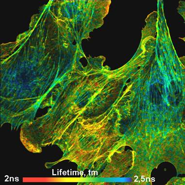
Fig. 1: BPAE cells stained with Alexa 488 and Mito Tracker red. FLIM
image, recorded by LSM 710 In Tune system with bh HPM‑100‑40
GaAsP detector and bh Simple-Tau 150 FLIM system.
The image shows BPAE cells (Molecular
Probes sample) stained with Alexa 488 and Mito Tracker red. The FLIM data were
recorded by a bh Simple-Tau 150 FLIM system [1, 2, 3] with a bh HPM‑100‑40
GaAsP hybrid detector [3, 4]. An image format of 512x512 pixels and 256 time
channels was used. The optical parameters were: 63x NA=1.4 oil immersion lens,
pinhole 1 AU, excitation wavelength 490 nm, laser power 1%, detection
wavelength from 505 nm to 700 nm. The lifetime displayed is the
amplitude-weighted average of a double-exponential fit to the data.
The high efficiency of the optics together
with the high efficiency of the bh GaAsP hybrid detector, tuneability and plenty
of laser power available allows one to use the system with diffraction-limited
pinhole sizes. It is thus easy to obtain diffraction-limited image quality.
More important, diffraction-limited pinhole size yields near-ideal axial
resolution. Axial resolution is extraordinarily important to FLIM.
Contamination from other focal planes adds unwanted decay components to the
recorded decay functions. The lifetime accuracy can thus be seriously impaired
by out-of-focus light, especially if the decay functions in the focal plane are
multi-exponential themselves.
Due to the high optical quality and the
absence of background signals the In Tune system performs surprisingly
well even for thick samples. Fig. 2 shows autofluorescence FLIM images of a pig
skin sample. The left image is 10 µm, the right image 40 µm from the
top of the sample. Both images show the amplitude-weighted lifetime, tm, and
the ratio of the amplitudes, a1/a2, of the lifetime components of a double
exponential decay model.
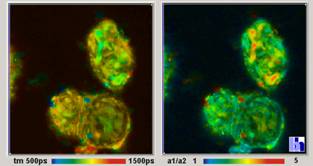
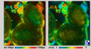
Fig. 2: Pig skin sample, autofluorescence. Double-exponential fit,
amplitude-weighted mean lifetime and amplitude ratio. 10 µm (left) and
40 µm (right) from the surface of the skin. HPM‑100‑40 hybrid
detector, SPC‑150 FLIM module.
The multi-exponential decay parameters of
autofluorescence decay, in particular amplitude ratios, have been shown to bear
information about the metabolic state of the tissue [5, 6]. Fig. 2 shows that
the In Tune system not only works in thick tissue, but also delivers
biologically relevant information. The fluorescence decay in a selected pixel
of Fig. 2, left, is shown in Fig. 3. The data quality is so good that even a
triple-exponential analysis delivers reasonable results.
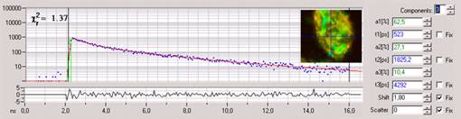
Fig. 3:
Fluorescence decay in selected spot of Fig. 2. Triple-exponential analysis.
The high optical quality, the spectral
purity of the excitation, and the high efficiency of the optics and the hybrid
detector make the LSM 710 In Tune FLIM also a good FCS system. An example
is shown in Fig. 4. Because the HPM‑100 hybrid detectors are free of
afterpulsing clean FCS is obtained down to 100 ns is obtained from a
single detector [3, 4].
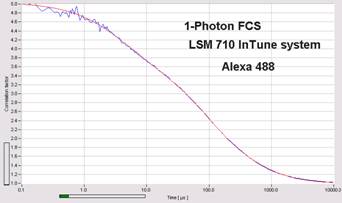
Fig. 4: FSC curve of Alexa 488 in water. bh HPM‑100‑40 GaAsP
hybrid detector with bh Simple-Tau 150 FLIM system.
The clean response of the HPM‑100‑40
detector [4] in combination with the short pulse width of the In Tune
laser makes gated FCS efficient in suppressing Raman signals. An example is
shown in Fig. 5. The left recording is contaminated by Raman emission of the
solvent. Because the Raman light has no fluctuations the correlation amplitude
obtained is too small. In the right recording, the Raman light has been gated
off by the SPC‑150 module of the FLIM system, and the correlation
amplitude is obtained correctly.
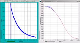
Fig. 5: Gated FCS. Left: Ungated recording. The signal is contaminated by
Raman emission from the solvent. Right: Raman component gated off. Decay curves
normalised.
Summary
The In Tune FLIM systems deliver FLIM data
at excellent sensitivity and spatial resolution. The systems deliver excellent
images both for single cells and tissue layers as deep as 40 µm. FCS is
obtained at high efficiency, and can be combined with time-gating to suppress
Raman signals. Unfortunately the high performance comes at a price: The
tunability range of the In Tune laser ends at 488 nm. The In Tune system therefore
cannot be used to excite the cyan fluorescent proteins. These are currently the
most frequently used donors in FLIM-based FRET measurements. Fortunately, a bh
BDL SMC picosecond diode laser is available for the LSM 710 [3]. We
recommend to add this laser to make FLIM at shorter excitation wavelength
possible.
References
1.
W. Becker, Advanced time-correlated single-photon counting techniques. Springer, Berlin,
Heidelberg, New York, 2005
2. W. Becker, The bh TCSPC handbook. Becker & Hickl GmbH (2005),
www.becker-hickl.com
3.
Becker & Hickl GmbH, Modular FLIM
systems for Zeiss LSM 510 and LSM 710 laser scanning microscopes.
User handbook. Available on www.becker-hickl.com
4.
Becker & Hickl GmbH, The HPM‑100-40
hybrid detector. Application note, available on www.becker-hickl.com
5.
V. Ghukasyan, F.-J. Kao, Monitoring cellular
metabolism with fluorescence lifetime of reduced nicotinamide adenine
dinucleotide. J. Phys. Chem. (2009)
6.
M. C. Skala, K. M. Riching, D. K. Bird, A.
Dendron-Fitzpatrick, J. Eickhoff, K. W. Eliceiri, P. J. Keely, N. Ramanujam, In
vivo multiphoton fluorescence lifetime imaging of protein-bound and free
nicotinamide adenine dinucleotide in normal and precancerous epithelia. J.
Biomed. Opt. 12 02401-1 to 10 (2007)
