4.4 ps IRF width of TCSPC with an NbN
Superconducting Nanowire Single Photon Detector
Wolfgang Becker, Becker & Hickl Gmbh
Jens Breffke, Boston Electronics Corp.
Boris Korzh, Matthew Shaw, Jet Propulsion Laboratory,
California Insitute of Technology
Qing-Yuan Zhao, Karl Berggren, EECS Deptartment,
Massachussets Institute of Technology
Abstract: An NbN superconducting nanowire single
photon detector (SNSPD) was used to perform time-correlated single photon
counting (TCSPC) with an instrument response function (IRF) width of 4.4 ps
full width at half maximum (FWHM). The detector was developed collaboratively
by MIT and the Jet Propulsion Laboratory (JPL), and the TCSPC module was a
modified BH SPC-150 NX device. We demonstrate the resolution of the system
by recording the fluorescence decay of IR 1061 (an infrared dye). The
fluorescence lifetime of IR 1061 dissolved in dichloromethane was
determined to be 43.7 ps.
Detector
Superconducting nanowire single photon
detectors (SNSPDs) are high-performance single-photon counting detectors consisting
of an ultra-thin superconducting strip biased by a DC current close the
critical value. The absorption of a photon in the superconducting wire leads to
a loss of superconductivity in the wire, increasing the resistance by several
kilohms and shunting the bias current into a readout amplifier. More
information about SNSPD technology and the detection mechanism can be found in
[1]. SNSPDs have recently been demonstrated with a time resolution of 2.7 ps
FWHM at 400 nm, and less than 5 ps FWHM time resolution at 1550 nm [2]. These
ultra-low-jitter devices consist of NbN nanobridges which are 7 nm thick, 80 nm
wide, and 5 µm long. While these proof-of-concept devices have negligble active
area, it is predicted that larger devices which can be efficiently coupled to
single-mode and multi-mode beams can still be engineered to have comparably low
timing jitter by using a differential readout technique [3].
The detector described in reference [2] was
used in a TCSPC experiment performed at JPL. In this experiment, the detector
was cooled below 1 K using a closed-cycle Helium-4 sorption refrigerator, and
read out using a low-noise SiGe cryogenic amplifier at the 4 K stage (Cosmic
Microwave Technology CITLF1). The light from the experiment was coupled onto
the detector through free-space windows and filters, using a lens located
outside the cryostat, thereby avoiding the temporal dispersion introduced by
fiber optics. SNSPDs have been used in the past for demonstrations of FCS [4]
and singlet oxygen detection [5], but the detector-TCSPC combinations used in
these experiments had timing jitter that limited the IRF width to 70 ps. Sub-30-ps
IRF width of an SNSPD with a BH SPC-150 TCSPC device has been achieved by Toussaint
et al. [8], 17 ps IRF width with a SNSPD from SCONTEL, Moscow [9] and a BH SPC-150N device [6].
TCSPC Device
For recording the shape of the light pulses
we used a BH SPC-150 NX module [7]. The SPC-150 NX has input
discriminators with 5 GHz bandwidth. Noise-optimized TAC-readout cricuitry
results in an electrical IRF width of 3.5 ps FWHM. The minimum time
channel width is about 400 fs. For the experiments described here, we
modified the module by increasing the TAC transfer ratio (time-to-voltage
ratio) by a factor of two. This yields a minimum time channel width of 204 ps,
and further reduces TAC readout noise. The electrical IRF of the module is shown
in Fig. 1, left. The IRF width is 3 ps FWHM.
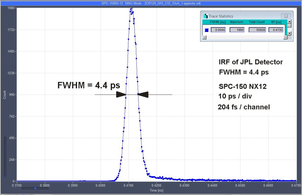
IRF Measurement
For the measurement of the optical IRF, we
used a Calmar Mendocino sub-ps mode-locked fiber laser. The laser delivers
pulses at 1064 nm wavelength, 50 MHz repetition rate, and an average power of
about 1 mW. The pulse width is 800 fs. Photons reflected off a solid
target were projected to the detector by a lens system. The light intensity was
adjusted by a variable ND filter in the laser beam path. The photon pulses from
the detector were connected to the 'CFD' (Start) input of the TCSPC module.
Timing reference pulses were generated by a fast photodiode. The pulses were
connected into the 'SYNC' (Stop) input of the TCSPC module. An IRF recorded
this way is shown in Fig. 1, right. The IRF width is 4.4 ps.
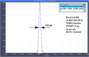
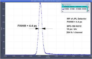
Fig. 1: Left: Electrical IRF of SPC-150 NX12 TCSPC module. Right: IRF
of detector, including laser pulse width and possible synchronization jitter.
Fluorescence Decay Measurement
Fluorescence decay measurements were
performed in the setup shown in Fig. 2. The dye solution was contained in a
cuvette. We used a 10-mm glass cuvette that had walls of 4 mm thickness on
two opposite sites. The horizontal cross section of the liquid volume is thus 2 mm
by 8 mm. The laser beam was focused into the cuvette by an IR apochromate,
L1. Fluorescence emitted by the dye solution is collected by another
apochromate, L2. The beam is collimated by L2, and focused on the detector by an
achromatic lens, L3. Two mirrors, M1 an M2, are used for alignment.
To avoid pulse broadening by transit time
effects it is important that the laser beam is focused correctly into the center
of the cuvette and that the fluorescence from exactly this spot is projected onto
the detector. The detector then forms a confocal system with the excited spot.
The effect is that out-of-focus detection is strongly suppressed. The
suppression of out-of-focus light in this setup is in fact so good that a clean
fluorescence signal is detected even without an emission filter.
Fig. 2: Optical
setup for fluorescence detection
For testing the setup we used a solution of
IR 1061, an IR dye from Sigma Aldrich. The fluorescence lifetime of IR 1061 is shorter
than 50 ps. With conventional fluorescence lifetime spectrometers the
fluorescence decay cannot be reliably resolved. A decay curve recorded with the
NbN detector described above is shown in Fig. 3. The time scale is 40 ps per
division, or 400 ps over the entire observation-time interval. The
resolution is so high that the curve almost looks like a
nanosecond-fluorescence decay recorded by a 'normal' lifetime spectrometer.
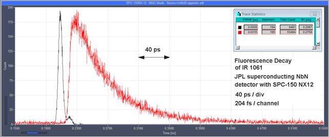
Fig. 3:
Fluorescence decay of IR 1061. Black: IRF, measured in diluted milk. Red:
Fluorescence
At the high time resolution of the system,
transit time differences at the time scale of one ps and below become visible.
For example, the fluorescence in Fig. 3 is visibly shifted against the IRF. The
reason is that the IRF was determined in water with a refractive index of 1.33,
but the dye was dissolved in dichloromethane, with a refractive index of 1.424.
The transit times of the laser pulses over the path length of 5 mm within
the solvent are 22.17 ps and 23.73 ps, respectively.
The difference is 1.56 ps, which becomes visible in data with such high
time resolution. Moreover, the IRF width measured in the cuvette is
5.8 ps, compared to 4.5 ps for the detection system itself. The
broadening is explained by the fact that the fluorescence is detected from a
non-zero sample volume: Already a longitudinal spread of 0.25 mm accounts
to a pulse broadening of about 1 ps.
A single-exponential fit with bh SPCImage
data analysis software is shown in Fig. 4. The fit delivers a fluorescence
lifetime of 43.7 ps.
Fig. 4: Single-exponential
fit with bh SPCImage data analysis software. Green curve shows IRF, blue dots
data points of the fluorescence decay curve, red curve fit with
single-exponential decay model. The fluorescence lifetime is 43.7 ps.
SPCImage includes a shift if the IRF
against the decay data in the fit procedure. The fit of the IRF shift delivers
a narrow χ2 minimum because the fluorescence decay time is more
than 7 times longer than the IRF width. It thus does not noticeably impair the
fit accuracy. Under these conditions, a single-exponential fit with SPCImage
delivers a (relative) standard deviation close to the theoretical value of  [71]. The
standard deviation of the lifetime obtained from the IR 1061 data can thus
be estimated from the number of photons in the photon trace. With 48.000
photons in the trace, the relative standard deviation is expected to be about
0.5 %.
[71]. The
standard deviation of the lifetime obtained from the IR 1061 data can thus
be estimated from the number of photons in the photon trace. With 48.000
photons in the trace, the relative standard deviation is expected to be about
0.5 %.
Summary
To the best of our knowledge, the setup presented
in this application note is the fastest TCSPC system ever described. With an
IRF width of 4.4 ps, it easily resolves fluorescence lifetimes in the
range of 50 ps and below. It also has the potential of resolving
ultra-fast effects like protonation reactions or solvent-relaxation effects.
Another application may be scanning of the surface profile of an object. The
time resolution of 4.4 ps does, in principle, resolve depth differences of
1.3 mm with a single detected photon in each pixel. With 1000 photons per
pixel the depth resolution would go down to about 40 µm. A frequent
concern with NbN detectors is the low sensitivity due to the small detector active
area. Although the quantum efficiency of the detector is close to one for the
microbridge detector used in this experiment, only a fraction of the photons
can be focused on the active area. The results presented above show the fluorescence
decay functions are well detectable even with moderate focusing. Replacing the
100 mm lens in front of the cryostat by a 20 mm lens inside it would
increase the efficiency by a factor of 25, which should be sufficient for a
wide range of fluorescence experiments. Furthermore, it is anticipated that
efficient, large-area SNSPDs with sub-5 ps time resolution can be engineered by
using a differential readout to remove the geometric jitter [3].
References
1.
C.M. Natarajan, M.G. Tanner, and R.H. Hadfield,
Superconducting nanowire single-photon detectors: physics and applications.
Superconductor Science and Technology 25(6), 063001 (2012)
2. B.A. Korzh et al, Demonstrating sub-3 ps temporal resolution in a
superconducting nanowire single-photon detector. ArXiv:1804.06839 (2018)
3. N. Calandri, Q-Y Zhao, D. Zhu, A. Dane, and K.K.Berggren, Superconducting nanowire detector jitters limited by detector
geometry. Appl. Phys. Lett., 109,
152601 (2016)
4. T. Yamashita et al, Fluorescence correlation spectroscopy with
visible-wavelength superconducting nanowire single-photon detector, Opt. Exp.
22(23), 28783 (2014)
5. N. Gemmell et al, Singlet oxygen luminescence detection with a
fiber-coupled superconducting nanowire single-photon detector. Opt. Exp. 21(4),
5005 (2013)
6. V. Shcheslavskiy, P. Morozov, A. Divochiy, Yu. Vakhtomin, K.
Smirnov, W. Becker, Ultrafast time measurements by time-correlated single
photon counting coupled with superconducting single photon detector, Rev. Sci.
Instrum. 053117 (2016)
7. W. Becker, The bh TCSPC Handbook, 6th ed., Becker & Hickl GmbH
(2015). Electronic version available on www.becker-hickl.com
8. J. Toussaint, S. Dochow, I. Latka, A. Lukic, T. May, H.-G. Meyer, K. Ilin, M. Siegel, J. Popp,
Proof of concept of fiber dispersed Raman spectroscopy using superconducting
nanowire single-photon detectors. Opt. Expr. 23, 5078-5090 (2015)
9. A. Verevkin, A. Pearlman, W. Slysz, J. Zhang, M. Currie, A. Korneev,
G. Chulkova, O. Okunev, P. Kouminov, K, Smirnov, B. Voronov, G. Goltsman, R.
Sobolewski, Untrafast superconducting single-photon detectors for
near-infrared-wavelength quantum communications. J. Mod. Op. 51, 1447-1458
(2004)
Acknowledgements
Part of this
research was carried out at the Jet Propulsion Laboratory, California Institute
of Technology, under a contract with the National Aeronautics and Space
Administration.
Contact:
Wolfgang Becker, Becker & Hickl GmbH, Berlin,
Germany, becker@becker-hickl.com
Boris Korzh, Jet Propulsion Laboratory,
California Institute of Technology, Pasadena, CA, boris.a.korzh@jpl.nasa.gov
Jens Breffke, Boston Electronics Corp., Boston, MA, jens@boselec.com