Fast-Acquisition Multiphoton FLIM with the Zeiss LSM 880 NLO
Abstract: This application note demonstrates the performance of
the bh FASTAC (fast acquisition) FLIM system in combination with the Zeiss LSM
880 NLO multiphoton laser scanning microscopes. The system is based on fast
distribution of the photon pulses of a PMH-100 hybrid detector into four
parallel TCSPC FLIM channels. The principle strongly reduces counting loss and
pile up effects. If the sample allows, images can be recorded in excess of
10 MHz count rate, and at acquisition times down to the minimum frame
times of the Zeiss LSM 880. Importantly, the system makes no compromises in
terms of time resolution, time channel width, time channel number, or pixel
number. The IRF width with fast detectors is less than 25 ps FWHM, and the
temporal data are recorded with typically 1024 time channels per pixel. The
time channel width can be made as small as 0.8 ps.
Principle
The system is based on the bh FASTAC fast
acquisition FLIM system [10, 11]. The FASTAC system uses a single detector the photon
pulses of which are distributed into four parallel TCSPC modules, see Fig. 1. The data of the four modules are combined into a
single FLIM data set. The TCSPC modules are running the normal bh FLIM process
[1, 2, 3]. Consequently, there is no need to trade time
resolution or time channel width against acquisition speed. The IRF width with
fast hybrid detectors is less than 25 ps (full width at half maximum) [7, 8, 10], the time channel width can be made shorter than 1
ps [1], and the number of time channels is large enough for
multi-exponential decay analysis [1, 8, 13]. Moreover, the multi-dimensional features of the bh
FLIM technique, such as spatial and temporal mosaic FLIM [1, 3], FLITS [4] or simultaneous FLIM / PLIM [5] remain available.
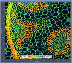
Fig. 1: Left: Principle of the bh FASTAC FLIM system. Right: Image of a convallaria sample, 512x512 pixels,
recorded in 4 seconds.
System Architecture
For the results presented in this note we
used an LSM 880 NLO multiphoton microscope with non-descanned detection. The
multiphoton version was chosen because most FLIM experiments are performed on
live cells and tissue [3]. The results can, however, extrapolated to one-photon
systems with ps diode laser excitation [13].
A HPM-100-06 hybrid detector [7] was attached to the NDD port of the microscope via
the usual Zeiss T adapter. The output pulses of the detector were connected to
a PHDIS photon distribution module [10]; the pulses from the four outputs of the PHDIS were
connected to four SPC-150N TCSPC / FLIM modules [1]. The data acquisition in the FLIM system was
synchronised with the LSM 880 via the usual pixel, line, and frame clock
signals [13]. The LSM 880 was controlled by the Zeiss ZEN
software, the FLIM system by bh SPCM software, version 9.78. The parameters for
general FLIM acquisition, TCSPC-channel combination, and online lifetime
display were chosen as described in [9], [12], and [13]. Multi-exponential data analysis was performed by bh
SPCIMage, version 7.1 [1, 13].
Results
Megapixel Images Obtained at Short Acquisition Time
In 2014 bh introduced Megapixel FLIM with
pixel formats of 1024 x 1024 and more, without compromising the temporal
resolution [6]. Fig.
2 shows that Megapixel FLIM is also available for the
FASTAC FLIM system. The figure shows a 1024 x 1024 pixel, 1024 time-channel
image of a BPAE (bovine pulmonary artery) cell sample (stained with DAPI, Alexa
488 and Mito Tracker Red). The excitation wavelength was 800 nm. The image was
recorded in 10 seconds. With normal FLIM systems several minutes are required
to record lifetime images of similar pixel numbers and similar signal-to-noise
ratio.

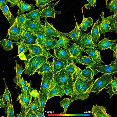
Fig. 2: FLIM of a BPAE sample. 1024x1024 pixels, 1024 time channels per
pixel. Acquisition time 10 seconds
Medium-Size Images
Another lifetime image of a BPAE sample,
recorded with 512 x 512 pixels and 1024 time channels, is shown in Fig. 3. The acquisition time was 10 seconds. A decay curve
from an 8x8 pixel region in the centre of the image is shown on the right.
Please note the fast rise of the fluorescence signal, which is a result of the
short IRF width of the detector / TCSPC combination [7].
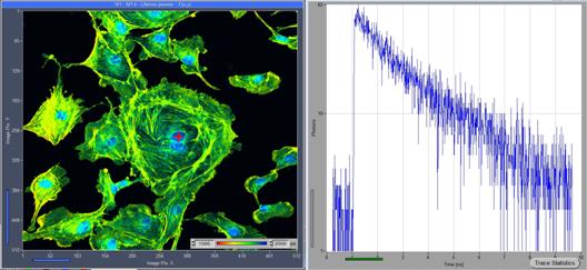
Fig. 3: FLIM image of a BPAE sample, 512 x512 pixels, 1024 time channels,
excitation wavelength 800 nm. Acquisition time 10 seconds. Left: Lifetime
image, created by online lifetime function of SPCM. Right: Decay curve from 8x8
pixel area around the spot marked in the image. Note the fast rise of the
fluorescence, which is a result of the extremely fast IRF of the system.
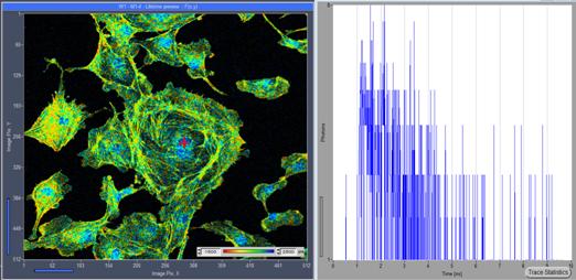
Fig. 4: Same
sample and sample area as in Fig.
3, but image recorded within 0.6 seconds
Fig.
4 shows an image of the same area of the same sample,
but recorded within a single frame of 0.6 seconds. Necessarily, the image gets
more noisy than the 10-second image. Nevertheless, the online-lifetime
calculation algorithm of SPCM [9] clearly reproduces the lifetime variation between
different regions of the cells.
The images shown in Fig. 2, Fig.
3 and Fig.
4 were recorded at an excitation power of 4% of the
available laser power. At this power, the BPAE sample delivered a count rate of
about 4 MHz averaged over the entire sample. The peak count rate probably
reached 10 MHz. This is significantly less than the FASTAC system can
process. In principle, the count rate and thus the signal-to-noise ratio could
be increased by increasing the laser power. However, laser power levels of 5%
and more caused rapid photobleaching and even photodamage in the sample.
Higher count rates can be obtained from
samples which contain higher concentrations of fluorophores. A typical representative
is the convallaria sample (stained
with acridine orange) often used for microscope demonstrations. Fig. 5 shows two images of a convallaria sample from Lieder.
Both images have 512 x 512 pixels and 1024 time channels per pixel. The left
image was recorded in 0.6 seconds, the right image in 4 seconds. Despite
the short acquisition time, the lifetime noise in the 0.6-second image is
barely visible. In the four-second image it is not visible at all.
Fig. 5: Convallaria sample, 512 x 512 pixels, 1024 time channels. Left:
Acquisition time 0.6 seconds. Right: Acquisition time 4 seconds. Images
calculated by online-FLIM algorithm of SPCM.
The data quality of the 4-second image is
high enough for double-exponential decay analysis. Fig. 6 shows images of the amplitude-weighted lifetime, tm,
and the amplitude-ratio, a1/a2. Lifetime images of the fast and the slow decay
component of the double-exponential decay are shown in Fig. 7.
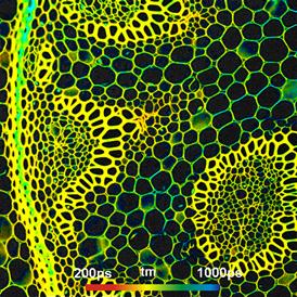
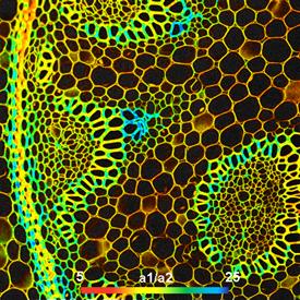
Fig. 6: Same data as in Fig.
5, right. Double-exponential FLIM analysis by bh
SPCImage. Amplitude-weighted lifetime, tm, (left) and amplitude ratio of decay
components, a1/a2 (right)
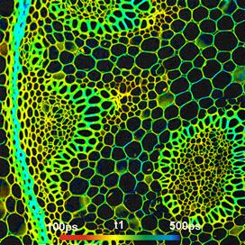
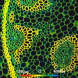
Fig. 7: Lifetime
image of fast decay component, t1, (left) and lifetime image of slow decay
component, t2, (right)
In many instances, images of the decay
times and the amplitudes of the components give a far better insight into
molecular parameters of a sample than simple lifetime images. It is therefore
important that a fast FLIM system delivers such data, and that it does so at
high precision. High time resolution (< 25ps FWHM IRF width with fast
detectors) substantially improves the accuracy of multi-exponential decay
analysis, as has been shown recently for metabolic FLIM [8].
Small Images
Small images (with small number of pixels)
can be recorded with even shorter acquisition time. One reason is that, at a
given count rate, the number of photons per pixel increases with decreasing
pixel number. Correspondingly, the signal-to-noise ratio of the lifetime
increases [3]. Another reason is that an optical scanner scans a
smaller image in a shorter period of time. The minimum frame time of the
scanner depends on the number of lines, not on the number of pixels along the
line. Therefore, if a scanner is operated at maximum speed and the frame format
(Pixels X × Pixels Y) is reduced the pixel dwell
time increases. In the FLIM
recording, the number of photons per pixel increases and so does the
signal-to-noise ratio of the lifetime. A few examples are shown in Fig. 8.
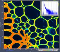
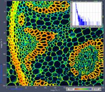
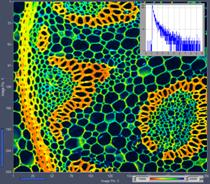
Fig. 8: FLIM data format 256 x 256 pixels, 1024 time channels.
Left to right: Acquisition time 0.16 s (zoom to obtain short frame time),
0.5 s, 2.5 s. Insert: Decay data in 5x5 pixel area around the cursor
position. Online lifetime display of SPCM.
Conclusions
The combination of the Zeiss LSM 880
NLO with the bh FASTAC FLIM system records FLIM images at acquisition times
down to the fastest frame rates of the LSM 880 scanner. There is no
compromise in terms of time-channel width, time-channel number, or pixel
numbers. The system can therefore be used both for fast-acquisition FLIM
applications and for precision FLIM. It should be noted, however, that fast
acquisition is only possible if the sample is able to feed the system with a
sufficiently high photon rate [11]. This is certainly the case for samples that contain
high amounts of bright fluorophores. It may not always be the case in molecular
imaging experiments, metabolic FLIM, or other applications where the fluorophores
are linked to highly specific targets within the cells. However, even under
sample-limited conditions the results are at least as good as with a standard
bh FLIM system.
References
1.
W. Becker, The bh TCSPC
handbook. Becker & Hickl GmbH, 7th ed. (2017). Available on
www.becker-hickl.com
2.
W. Becker, Advanced
time-correlated single photon counting techniques. Springer, Berlin, Heidelberg, New York
(2005)
3.
W. Becker (ed.), Advanced
time-correlated single photon counting applications. Springer, Berlin, Heidelberg, New York
(2015)
4.
W. Becker, V. Shcheslavkiy, S.
Frere, I. Slutsky, Spatially Resolved Recording of Transient
Fluorescence-Lifetime Effects by Line-Scanning TCSPC. Microsc. Res. Techn. 77,
216-224 (2014)
5.
Becker & Hickl GmbH,
Simultaneous Phosphorescence and Fluorescence Lifetime Imaging by
Multi-Dimensional TCSPC and Multi-Pulse Excitation. Application note, available
on www.becker-hickl.com
6.
Becker & Hickl GmbH, Megapixel
FLIM with bh TCSPC Modules: The New SPCM 64-bit Software. Application note,
available on www.becker-hickl.com
7.
Becker & Hickl GmbH,
Sub-20ps IRF Width from Hybrid Detectors and MCP-PMTs. Application note,
available on www.becker-hickl.com
8.
Becker & Hickl GmbH,
Ultra-fast HPM Detectors Improve NAD(P)H FLIM. Application note, available on
www.becker-hickl.com
9.
Becker & Hickl GmbH, SPCM
Software Runs Online-FLIM at 10 Images per Second. Application note, available
on www.becker-hickl.com
10.
Becker & Hickl GmbH, Fast-Acquisition
TCSPC FLIM System with sub-25 ps IRF Width. Application note, available on
www.becker-hickl.com
11.
Becker & Hickl GmbH, Fast-Acquisition
TCSPC FLIM: What are the Options? Application note, available on
www.becker-hickl.com
12.
Becker & Hickl GmbH, New
SPCM Version 9.78 comes with new software functions. Application note, available
on www.becker-hickl.com
13.
Becker & Hickl GmbH,
FLIM Systems for Zeiss LSM 710 / 780 / 880 Family Laser Scanning Microscopes.
User Handbook, 7th ed. (2017). Available on www.becker-hickl.com
Contact:
Wolfgang Becker
Becker & Hickl GmbH
Berlin, Germany
Email: becker@becker-hickl.com
info@becker-hickl.com