FLIM of Macroscopic
Objects: Imaging in the Primary Image Plane of the DCS‑120 Scanner
Wolfgang Becker, Becker & Hickl GmbH
Abstract: Fluorescence lifetime images of macroscopic sample as large as
15 mm can be recorded in the primary focal plane of the DCS‑120
confocal scanner. Optical resolution is in the range of 15 µm, acquisition
times range from a few seconds for low pixel numbers to several minutes for
high-resolution images.
Principle
Multidimensional TCSPC is a powerful
technique to obtain fluorescence lifetime images of microscopic samples in
laser scanning microscopes [1, 2]. A laser beam is sent down the beam path of
the microscope, and scanned around a pivot point located in the principle plane
of the microscope lens. The fluorescence light is collected back through the
microscope lens, separated from the excitation by a dichroic mirror, and
detected in one or several wavelength intervals or polarisation channels. Depth
resolution or optical sectioning is obtained by confocal detection [3] or
multiphoton excitation [4, 5].
Images obtained in microscopes usually
cover image areas of no more than 1 x 1 mm. Larger objects can,
in principle, be scanned by placing them in the intermediate image plane of a
confocal scanner. The optical principle is shown in Fig. 1. The figure shows
the internal beam path of the bh DCS-120 scanner, but the principle shown essentially
applies to other confocal scanners as well.

Fig. 1:
Principle of the DCS-120 scanner for imaging macroscopic objects (simplified)
The laser beam is scanned by two
fast-moving galvanometer mirrors. The scan lens focuses the laser beam into an
image plane shortly in front of the scanner. When the scanner is used with a
microscope, this image plane coincides with the upper image plane of the
microscope. For scanning large samples, the image plane of the scan lens is
brought in coincidence with the sample surface. As the galvanometer mirrors change
the beam angle the laser focus scans across the sample. Fluorescence light
excited in the sample is collimated by the scan lens, de-scanned by the
galvanometer mirrors, and separated from the excitation light by the main
dichroic beamsplitter. The fluorescence beam is further split into two spectral
or polarisation components, and focused into pinholes. Light passing the
pinholes is sent to the detectors. For more details please see [3].
Results
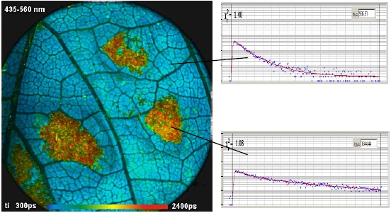
Fig. 2 shows two examples of images
recorded in the primary image plane of the DCS‑120 scanner.
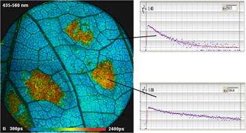
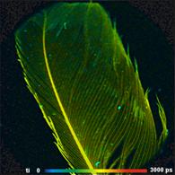
Fig. 2: FLIM in the primary image plane of the DCS-120 scanner, excitation
405nm, scan 512 x 512 pixels. Left: Leaf with a fungus infection. Middle:
Decay functions of healthy and infected areas. Right: Feather of a songbird.
Image area and Resolution
The maximum diameter of the image area in
the primary image plane of the scanner is about 15 mm. Smaller areas can
be scanned by using the Zoom function of the scanner, see [3]. The size of
the laser spot in the image plane is about 15 µm. The resolution in the
detection path is even higher because the numerical aperture in the focus of
the scan lens is larger than for the laser beam. That means, full-size images
could, in principle, be reasonably scanned with at least 1024 x 1024
pixels, with moderate oversampling even with 2048 x 2048 pixels.
Collection efficiency
The numerical aperture of the detection
beam path is given by the beam path diameter (about 3 mm) and the focal
length of the scan lens (40 mm). The collection efficiency is thus considerably
lower than in combination with a microscope. However, macroscopic imaging can
use much higher laser power, which compensates for low collection efficiency.
Even weakly fluorescent samples, such as human skin, or the feather shown in Fig.
2, easily deliver count rates on the order of several 100,000 counts per
second.
Acquisition times
The acquisition time depends on the number
of pixels in the image and the requirements to the accuracy of the lifetimes [2,
3]. The images shown above were recorded within about one minute. For images
with 128 x 128 pixels the same lifetime accuracy is obtained in less
than 10 seconds.
References
1. W. Becker, Advanced time-correlated single-photon counting techniques. Springer,
Berlin, Heidelberg, New York, 2005
2.
W. Becker, The bh TCSPC handbook. 4th edition,
Becker & Hickl GmbH (2008), available on www.becker-hickl.com
3. Becker & Hickl GmbH, DCS-120 Confocal Scanning FLIM Systems. User
handbook. www.becker-hickl.com
4. Becker & Hickl GmbH, Modular FLIM systems for Zeiss
LSM 510 and LSM 710 laser scanning microscopes. User handbook,
available on www.becker-hickl.com
5. Becker & Hickl GmbH, Non-Descanned FLIM Detection in Multiphoton
Microscopes. Application note, available on www.becker-hickl.com