FLIM Systems for Zeiss LSM 710 Record Z Stacks
Abstract.
The bh FLIM systems for the Zeiss LSM 710 and LSM 710 NLO microscopes
[5] are able to record Z stacks of FLIM images. The LSM 710 subsequently
scans a number of Z planes. Synchronisation of the FLIM recording with the Z
scan is obtained via a z-plane trigger from the microscope. The FLIM system
starts a FLIM acquisition for each plane trigger, and repeats the acquisition
for the desired number of Z planes. After each plane the data are saved into a
file. The result is a sequence of FLIM data files for consecutive planes of the
sample.
Introduction
The recording of Z stacks of images, or Z
scanning has been early introduced into confocal and multiphoton laser scanning
microscopes [11]. The microscope scans one image plane, saves the image data,
changes the depth of the focus, and scans another plane. The process is
continued until the desired number of planes have been scanned, see Fig. 1.
Ideally, a sufficiently large number of closely spaced z planes would be
scanned to allow the three-dimensional structure of the sample to be
reconstructed.

Fig. 1: Z
stack of algae, recorded by bh FLIM system, intensity images
Z stack recording faces two major problems.
First, the maximum depth in the sample from which a reasonable image can be
obtained is limited by absorption, scattering, refractive-index mismatch and
refractive index inhomogeneity. Second, scanning a large number of planes
exposes the sample to a high excitation dose. Photobleaching is therefore a severe
limitation. Both problems are mitigated by using two-photon excitation.
Absorption at NIR wavelengths is usually low, and scattered excitation light
can be collected by non-descanned detection (NDD) [8]. Different than for
one-photon excitation, photobleaching is restricted to the focal plane.
Although two-photon excitation causes increased photobleaching in the focal
plane [9] the total amount of bleaching for a large number of Z planes is
usually lower than for one-photon excitation [10].
Despite of these improvements,
photobleaching has been a severe obstacle to the introduction of Z stack FLIM
in the past 10 years. Although FLIM data are obtained from a given number of
photons at the same accuracy as intensity data FLIM usually requires a larger
number of photons per pixel [1, 2]. The reason is that the requirements to the
lifetime accuracy in FLIM experiments are normally higher than to the intensity
accuracy in steady-state experiments. Moreover, a moderate amount of
photobleaching can usually be tolerated for the reconstruction of spatial
structures. In FLIM, however, photobleaching can cause unpredictable lifetime
changes. Another obstacle to Z stack FLIM has been the synchronisation with the
Z stepping of the microscope. The FLIM acquisition within one plane is
synchronised via pixel, line, and frame clock pulses delivered by the
microscope [1, 6]. However, until recently, laser scanning microscopes did not
deliver a Z synchronisation pulse. Z scanning could therefore only be achieved
by actively controlling the microscope from the FLIM system [2]. This approach has been employed in the bh
DCS‑120 confocal scanning FLIM system [3, 4] but cannot be used in other
scanning microscopes because the Z control port is not accessible.
Z Stack FLIM with the Zeiss LSM 710
The Zeiss LSM 710 offers a number of
advantages over previous laser scanning microscopes. The NDD light path of the
multiphoton versions has been improved in efficiency, especially for scattered
photons from deep sample layers. The efficiency of the LSM 710 scan head
is near-perfect. The scan head has been equipped with an optional direct coupled
confocal port (DC port) which has a substantially higher efficiency than the
fibre output of the LSM 510. Moreover, the microscopes have been
complemented with a tuneable picosecond laser. This InTune laser allows the
optimum excitation wavelength to be selected for the fluorophores excited. All
these features have resulted in a substantial increase in the efficiency of
FLIM detection, and, consequently, in a reduction of photobleaching artefacts.
Another big leap in efficiency came with
the introduction of the HPM‑100‑40 hybrid detector modules by bh [7].
The efficiency of these detectors is 5 to 10 times better than for conventional
PMTs, and reaches or even surpasses the efficiency of single-photon avalanche
photodiodes (SPADs). Different than SPADs, the hybrid detectors have active
areas on the order of 7 mm2. This is a perfect match to the NDD
optics of the LSM 710, and thus provides unprecedented efficiency to
deep-tissue imaging [5]. At the DC port of confocal systems, the large area
avoids any alignment or focusing problems and collects light efficiently even
from large pinholes [5]. With the new detectors photobleaching no longer
prevents the use of Z scanning.
The last problem - the synchronisation of
the FLIM recording with the Z stepping - has been solved by introducing a
Z-plane trigger output into the Zeiss LSM 710 scan control system. The
principle of the synchronisation is shown in Fig. 2.
Fig. 2: Synchronisation
of the FLIM system with the Z stepping
Stack acquisition is achieved by using the
experiment trigger and autosave functions of the bh FLIM system in combination with
the Z stepping and frame accumulation function of the Zeiss LSM 710. The
LSM 710 real-time computer sends a plane trigger to the bh SPC‑150
modules when it starts to scan a plane. Each plane is scanned repetitively,
typically 4 to 16 times. The number of frames per plane, the frame time, the z
step width, and the number of Z planes to be scanned is defined in the ZEN
software of the LSM 710. For each plane, the FLIM system acquires FLIM data for
a defined collection time. The FLIM acquisition for the individual planes is
synchronised in the usual way via the frame clock, line clock, and pixel clock
pulses. When the collection time is over the FLIM system stops the acquisition,
saves the data into a file, and waits for the next plane trigger. The
acquisition continues for a number of Z planes defined by the cycles
parameter in the SPCM software of the FLIM system.
Typical results are shown in Fig. 3 and Fig.
4. Images of an (unstained) pig skin sample were recorded by a dual-channel
FLIM system in an LSM 710 NLO multiphoton microscope. HPM‑100‑40
hybrid detectors were used at the 0° and the 90° output of the NDD T-Adapter of
the LSM 710. A beamsplitter was used to record simultaneously images below
and above 480 nm. The excitation wavelength was 800 nm, the laser
power 2.4 %. A water apochromate 40 x, NA=1.1 was used; the scan area was 212 x 212
µm. The FLIM images of each plane were acquired at a resolution of 256 x 256
pixels and 256 time channels. The Z step interval chosen in the ZEN software
was 5.09 µm. The total scan time per plane was 25 seconds. The
collection time of the FLIM system was 20 seconds, i.e. 5 seconds were
spared for saving the data and waiting for the next plane trigger.
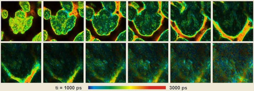
Fig. 3 shows the FLIM image in the
wavelength channel above 480 nm. The colour of images shows the average
(intensity-weighted) lifetime of a double-exponential fit to the decay data.
The intensity of the images was normalised to the intensity of the brightest
pixel.
Fig. 3: FLIM Z stack recorded at a pig skin sample, excitation at
800 nm, emission above 480 nm. Z step width 5.09 µm, scan area
212 x 212 µm. Images from 5 µm to about 60µm below the surface.
Colour represents average (intensity-weighted) lifetime of a double-exponential
fit. FLIM data format 256 x256 pixels, 256 time channels.
No decrease in count rate was observed
during the acquisition time of the individual Z planes. Moreover, after
returning to the first plane the count rate had not changed. The stability of
the count rate indicates that photobleaching was negligible.
Fig. 4 shows data recorded in the
wavelength interval below 480 nm. The data recorded in this interval
contain both fluorescence and SHG. The SHG signal can be extracted from these
data by selecting the signal component of infinitely short lifetime. Fig. 4
shows SHG images extracted from the FLIM data this way. The intensities were
normalised to the intensity in the brightest pixel.
Fig. 4: Z stack recorded at a pig skin sample, excitation at 800 nm,
emission below 480 nm, SHG signal, extracted by selecting photons in an
early time window. Z step width is 5.09 µm, scan area 212 x 212
µm. Images from 5 µm to about 60µm depth.
Summary
We have demonstrated Z stack FLIM by synchronising
the cycle and autosave functions of the bh FLIM systems with the Z stepping
function of the Zeiss LSM 710 NLO microscopes. By using non-descanned
detection and high-efficiency hybrid detectors autofluorescence and SHG images
of biological tissue are obtained at low excitation power, negligible
photobleaching, and within a reasonable acquisition time. Although Z stack FLIM
is especially useful in combination with two-photon excitation it is in no way
restricted to multiphoton systems. The FLIM system, the principle of
synchronisation, and even the system setup parameters are the same for
one-photon excitation by ps diode lasers and by the tuneable InTune laser of
the LSM 710 confocals.
References
1.
W. Becker, Advanced time-correlated single-photon counting techniques. Springer, Berlin,
Heidelberg, New York, 2005
2.
W. Becker, The bh TCSPC handbook, 3rd edition. Becker
& Hickl GmbH (2008), www.becker-hickl.com
3.
Becker & Hickl GmbH, DCS-120 Confocal
Scanning FLIM Systems, user handbook. www.becker-hickl.com
4.
Becker & Hickl GmbH, Recording Z Scans with
the DCS‑120 Confocal Scanning FLIM System. Application note, available on
www.becker-hickl.com
5.
Becker & Hickl GmbH, Modular FLIM
systems for Zeiss LSM 510 and LSM 710 laser scanning microscopes.
User handbook, available on www.becker-hickl.com
6. W. Becker, A. Bergmann, M.A. Hink, K. König, K. Benndorf, C. Biskup,
Fluorescence lifetime imaging by time-correlated single photon counting, Micr.
Res. Techn. 63, 58-66 (2004)
7.
Becker&Hickl GmbH, The HPM‑100-40 hybrid
detector. Application note, available on www.becker-hickl.com
8.
Becker & Hickl GmbH, Multiphoton NDD
FLIM. Application note, available on www.becker-hickl.com
9. P.S. Dittrich, P. Schwille, Photobleaching and stabilization of
fluorophores used for single-molecule analysis with one- and two-photon
excitation, Appl. Phys. B 73, 829-837 (2001)
10.
D.R. Drummond, N. Carter, R.A. Cross,
Multiphoton versus confocal high resolution z-sectioning of enhanced green
fluorescent nanotubules: Increased multiphoton photobleaching within the focal
plane can be compensated using a Pockels cell and dual widefield detectors. J.
Microsc. 206, 161-169 (2001)
11. J. Pawley
(ed.), Handbook of biological confocal microscopy, 2nd edn., Plenum Press, New
York (1995)