273 ps FWHM TCSPC Response with Hamamatsu H15620 NIR
PMT
Wolfgang
Becker, Taravat Saeb-Gilani, Alexander Jelzow
The new Hamamatsu
H15620 NIR PMT module delivers an instrument-response width of < 280 ps
FWHM with the bh SPC-150NX TCSPC module. The module fits smoothly into the bh
TCSPC systems. The supply voltage for the detector, the cooler current, and the
gain control voltage are available from a DCC-100 detector controller module.
In combination with a HFAC-26-1 preamplifier, the DCC-100 provides also for
overload shutdown. We demonstrate the performance of the detector at the
example of diffuse optical imaging experiments at a wavelength of 1300 nm.
TCSPC above 1000 nm
After more than 60 years of TCSPC
development [1, 2] the detection
of optical signals above a wavelength of 1000 nm is still a problem. InGaAs
SPADs work in the range from 900 to 1700 nm. They have high quantum
efficiencies and reasonably fast IRFs [3, 4] but the diameter of the active
area is on the order of only 20 µm. The detectors are therefore well
suited for systems that detect light from a diffraction-limited spot, such
confocal microscopes or micro-spectrometers [2, 4] but they are not efficient
in detecting light that emerges from a large area. An even wider wavelength
range is available from superconducting single-photon detectors (SSPDs). With single-nanowire
and meander-type SSPDs, bh TCSPC modules deliver IRF widths down to 4.4 ps
and 17 ps (fwhm), respectively [5, 6]. However, with active areas of 0.2 µm
x 2 µm and 4 x 4 µm, the detectors are extremely small, and
even difficult to handle in diffraction-limited optical systems. The use in
systems for diffuse optical experiments with biological tissue is therefore
impossible. PMTs with IR-sensitive cathodes do exist and offer large active
area. However, even with strong cooling the dark count rates are so high that
they present a substantial fraction of the saturated count rate of a TCSPC
device. This limits the dynamic range over which an optical waveform can be
recorded. Recently, Hamamatsu have addressed the problem by building a compact cooled
PMT module with an active area of 1.6 mm2. On the one hand,
this is small enough to keep the dark count rate at a reasonable level. On the
other hand, it is large enough to capture enough light from a diffusely emitting
object. This application note gives an overview on the essential parameters
reached by a TCSPC system with this detector.
TCSPC Performance of the Hamamatsu H15620 Detector
The Hamamatsu H15620 detector contains a
small PMT together with a high-voltage generator, a thermoelectric cooler, a
heat sink, and a cooling fan. The active area is 2 mm2. The
detector needs a +5V power supply, a 0...+0.9V gain control voltage, and a 1...4 A
current source for the thermoelectric cooler. These voltages and the thermoelectric-cooler
current are supplied by a bh DCC-100 detector controller module. The only
voltage which is not available from the DCC is the supply voltage for the
cooling fan. Together with a bh HFAC-26-1 26-dB preamplifier the DCC‑100
also provides for overload shutdown. The single-photon pulses from the
preamplifier have an amplitude of ‑100 to ‑400 mV. This is
compatible with the CFD input of the bh SPC series TCSPC / FLIM modules. The
connection diagram is shown in Fig. 1.
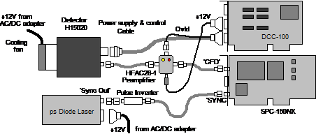
Fig. 1:
Connection diagram, TCSPC system with H15620 detector
IRF
Our test device was a H15620-45, with a
wavelength range from 950 nm to 1400 nm. For testing the IRF we used
a BDS-SM, 1300 nm picosecond diode laser. The laser beam was projected
through a set of ND filters to the photocathode of the detector. The beam
diameter was 4 mm, i.e. the entire active area was illuminated. The IRF
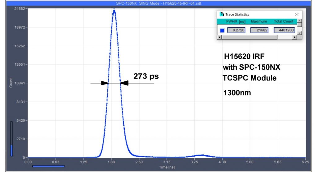
recorded this way is shown in Fig. 2.
Fig. 2: IRF of
the H15620-45 detector, recorded with SPC-150NX TCSPC module and BHL-150,
1300 nm picosecond diode laser. Full width at half maximum is 273 ps.
The IRF was recorded with a detector gain
control voltage of 0.9 V. The CFD parameters of the SPC-150 module were
optimised for best IRF shape and maximum detection efficiency. The CFD
threshold under these conditions was -200 mV, the CFD zero-cross level
+15 mV. No attempts were made to narrow the IRF on the expense of detection
efficiency.
With a full width at half maximum (fwhm) of
273 ps, the IRF is surprisingly fast - Hamamatsu specifies a typical value
of 400 ps. We do not know whether the fast IRF is a result of the
extremely fast discriminator of the SPC‑150 NX module or we just received
an extraordinarily fast detector. Please note also that the IRF width is for
the H15620-45. It may be different for the H15620-25 because of different
electron diffusion times in the photocathodes.
Dark Count Rate
As expected, the dark count rate depends on
the cooling current. The maximum cooling current for the H15620 is 5 A. The
DCC-100 detector controller delivers a maximum current of 2 A. With the
2 A from the DCC-100 we obtained a dark count rate of approximately 4000
counts per second. The ambient temperature was 25 °C, the cooling fan was
running at 12V. With a cooling current of 3 A (from an external power
supply) the dark count rate dropped to about 2500 counts per second.
Diffuse Optical Imaging Experiments
The expected application of the H15620 is
in diffuse optical tomography, or near-infrared spectroscopy (NIRS) and functional
near-infrared spectroscopy (fNIRS) [1, 2]. Scattering and absorption in biological
tissue decrease with increasing wavelength. Therefore, near-infrared light
penetrates relatively thick layers of tissue. The wavelength range around
1300 nm is of special interest because the absorption of water has a local
minimum at this wavelength.
A Distribution of Time of Flight (DTOF)
after propagation of 1300 nm light pulses through 25 mm of tissue
(the palm of a human hand) is shown in Fig. 3. The incident power was about 200 µW,
the pulse repetition rate 50 MHz.
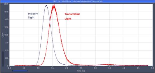
Fig. 3.
Distribution of time of flight (DTOF) after propagation of ps light pulses
through the palm of a human hand
Recording of DTOFs can be combined with imaging.
The principle is shown in Fig. 4. The laser beam is scanned over the object of
investigation by a galvanometer scanner, in our case a modified bh DCS-120 scan
head [2].
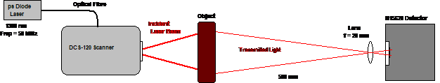
Fig. 4: DTOF
scan of a diffusely transmitting object, optical principle
The photons emerging from the distant side
of the object are transferred to a H15620-45 detector placed in a distance of
about 50 cm. A 20 mm lens in front of the detector projects a
de-magnified image of the object on the active area. The large distance is
necessary to obtain an image small enough to fit into the active area of the detector.
The photon pulses from the detector are recorded by the TCSPC module, which
builds up the distribution of the photons over the time in the laser pulse
period and the coordinates of the scan [2].
A scan of the palm of a human hand is shown
in Fig. 5. The left image shows the intensity of the transmitted light. Additional
information on scattering and absorption coefficients is obtained by moments of
the DTOFs in the individual pixels [7]. Therefore, in the right images the
first moment of the DTOFs has been calculated and an overlaid to the intensity
data by false colour.
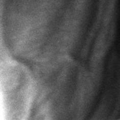

Fig. 5: DTOF scan of the palm of a human hand. Left: Intensity image.
Right: Combined intensity / propagation-time image. Colour shows the average
propagation time, yellow to green refers to 900 to 1100 ps.
Summary
The H15620 is fully capable of recording
optical waveforms in combination with the bh TCSPC devices. With its fast IRF,
relatively large area, and low dark count rate it is especially suitable for
detecting signals from diffusely emitting objects. Applications are
preferentially in the field of NIRS and fNIRs, including brain imaging and
optical mammography, in the optical window around 1300 nm. Other
applications may be in material sciences, especially in the investigation of
novel solar cell materials.
Acknowledgement
We thank Hamamatsu Photonics for providing
the H15620-45 test device.
References
1. W. Becker, Advanced
time-correlated single-photon counting techniques. Springer, Berlin,
Heidelberg, New York, 2005
2.
W. Becker, The bh TCSPC
handbook. 8th edition, Becker & Hickl GmbH (2019), available on
www.becker-hickl.com
3.
Becker & Hickl GmbH,
80 ps FWHM Instrument Response with ID230 InGaAs SPAD and SPC 150 TCSPC Module.
Application note, available on www.becker-hickl.com
4.
Becker & Hickl GmbH,
TCSPC at Wavelengths from 900 nm to 1700 nm. Application note, see
www.becker-hickl.com
5. W. Becker, J. Breffke, B. Korzh, M. Shaw, Q-Y. Zhao, K. Berggren, 4.4 ps IRF width of TCSPC
with an NbN Superconducting Nanowire Single Photon Detector. Application note,
available on www.beker-hick.com
6.
Becker & Hickl GmbH,
World Record in TCSPC Time Resolution: Combination of bh SPC-150NX with SCONTEL
NbN Detector yields 17.8 ps FWHM. Application note, see
www.becker-hickl.com
7.
A. Liebert, H. Wabnitz,
D. Grosenick, M. Möller, R.Macdonald, H. Rinneberg, Evaluation of optical
properties of highly scattering media by moments of distributions of times of
flight of photons, Appl. Opt. 42, 5785-5792 (2003)
Contact:
Wolfgang Becker
Becker & Hickl GmbH, Berlin, Germany
https:/www.becker-hickl.com
becker @becker-hickl.com