Principles
Table of Contents:
- Functional Brain Imaging with Time-Resolved Detection by TCSPC
- Advanced fNIRS Instrumentation for Dynamic Brain Imaging
Functional Brain Imaging with Time-Resolved Detection by TCSPC
NIRS (Near-Infrared Spectroscopy) techniques are able to record absorption and scattering changes in biological tissue down to a depth of several centimetres. Applied to the human head, the techniques can record dynamic changes in the time-of-flight distributions caused by the heart beat, by oxy- and deoxyhemoglobin changes during brain activity. The haemodynamic response to brain stimulation is on the time scale of a 100 ms to few seconds. Time-resolved detection by TCSPC provides improved separation of scattering and absorption, and better depth resolution than CW techniques. In particular, moment analysis and time-window analysis of the distributions of time of flight (DTOFs) provide a way to distinguish between intracerebral and extracerebral oxygenation changes.
Advanced fNIRS Instrumentation for Dynamic Brain Imaging
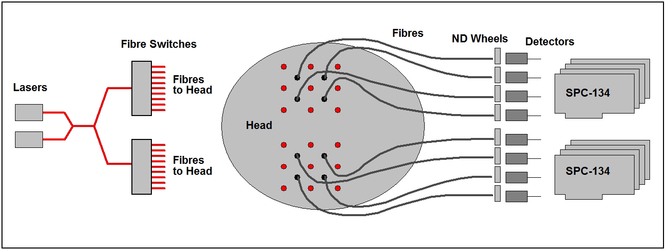
Instruments for functional brain imaging use similar setups as for static brain imaging. However, because fast sequential recording is required, the signals of the detectors are normally recorded by fully parallel TCSPC channels. The architecture of an instrument for dynamic brain imaging is shown in the figure below. The instrument uses two SPC-134 packages to obtain 8 parallel recording channels. The DTOFs at both hemispheres of the head are recorded simultaneously. Each hemisphere has 9 source fibres and 4 detection fibres attached. Two lasers of different wavelengths are multiplexed in time. In addition, a fibre switch periodically switches through the 9 source positions of each hemisphere. For every source position the DTOFs are acquired for 95 ms. One switching cycle through all 9 source positions is completed within 0.9 seconds. The sequences are recorded in the ‘Continuous Flow’ mode of SPC-130, SPC-130EM, or SPC-150N modules.
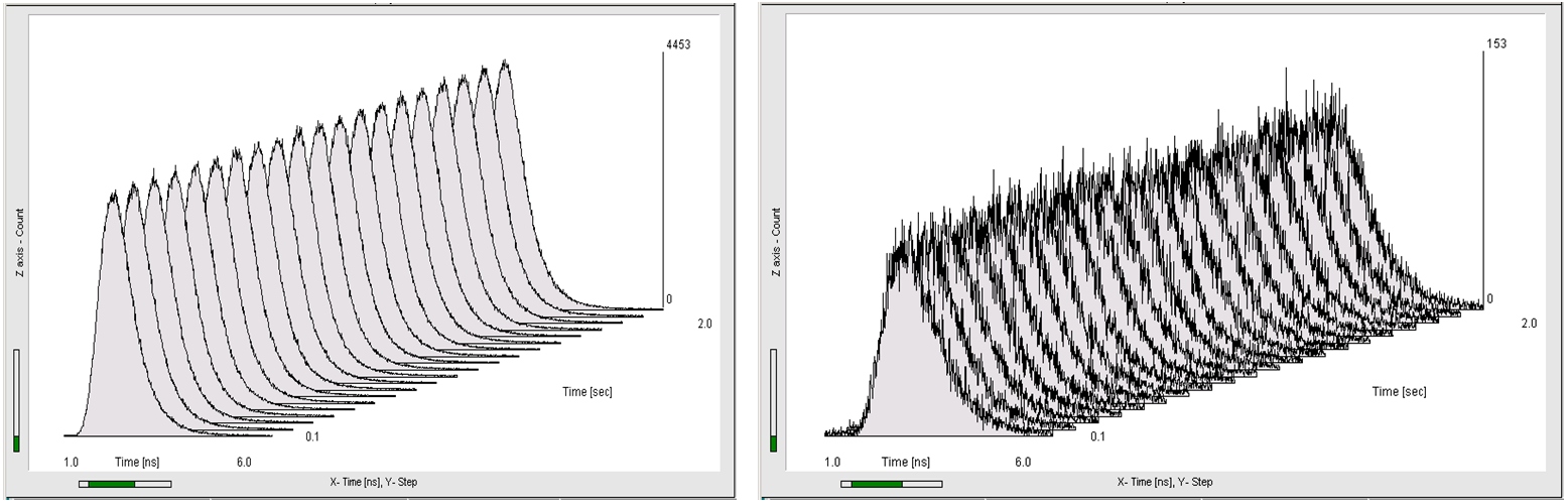
To give an impression of the of the recorded data, the figure below shows 20 steps of two different sequences of DTOFs, both recorded at a speed of 100 ms per curve.
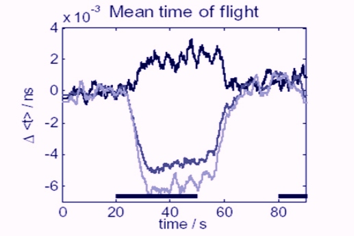
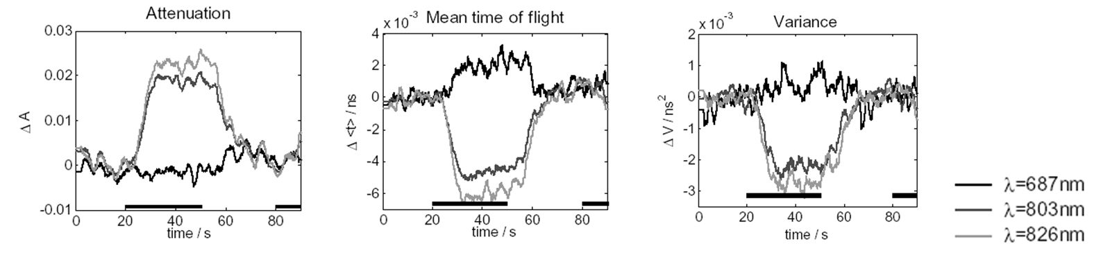
Variations of the optical properties in the brain are derived from the intensity and the first and second moments of the time-of-flight distributions. A typical result of a brain-stimulation experiment is shown in the figure below. As can be seen from the Mean Time of Flight curves, the variations are on the order of a few picoseconds. Therefore, a timing stability of the TCSPC modules of better than a picosecond is required to correctly record the response of the brain to the stimulation.
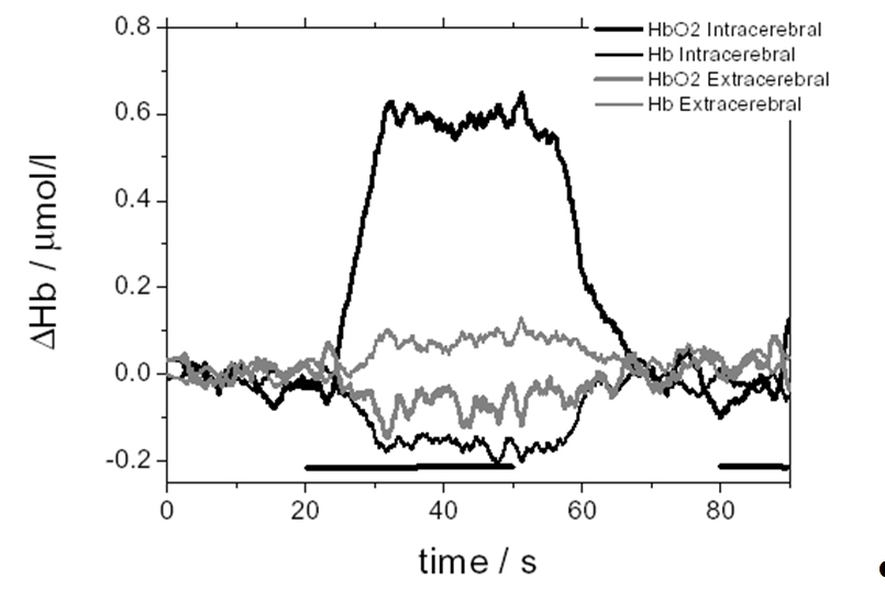
Depth-resolved intra- and extra-cerebral changes of the oxy- and deoxyhemoglobin concentrations calculated from the data at both wavelength, and all source and detector positions of one hemisphere are shown in the figure below.
For more information, related applications and references, please see:
The bh TCSPC Handbook, chapter ‘Diffuse Optical Tomography: DOT, NIRS and fNIRS’
References
References Related to fNIRS
More references in W. Becker, The bh TCSPC Handbook 7ed. (2017)
- Aletti, R. Re, V. Pace, D. Contini, E. Molteni, S. Cerutti, M. A. Bianchi, A. Torricelli, L. Spinelli, R. Cubeddu, G. Baselli, Deep and surface hemodynamic signal from functional time resolved transcranial near infrared spectroscopy compared to skin flowmotion. Comput Biol Med. 42(3), 282-289 (2012)
- Contini, A. Pifferi, L. Spinelli, A. Torricelli, R. Cubeddu, Multi-channel time-resolved tissue oximeter for functional imaging of the brain. IEEE T. Intrum. Meas. 55, 85-90 (2006)
- Contini, A. Torricelli, A. Pifferi, L. Spinelli, F. Paglia, R. Cubeddu, Multi-channel time-resolved system for functional near infrared spectroscopy. Opt. Expr. 14, 5418-5432 (2006)
- Durduran, G. Yu, M.G. Burnett, J.A. Detre, J.H. Greenberg, J. Wang, C. Zhou, A. Yodh, Diffuse optical measurement of blood flow, blood oxygenation, and metabolism in a human brain during sensorimotor cortex activation, Opt. Lett. 29, 1766-1768 (2004)
- A. Franceschini, S. Fantini, J.H. Thompson, J.P. Culver, D.A. Boas, Hemodynamic evoked response of the sensorimotor cortex measured noninvasively with near-infrared optical imaging, Psychophysiology 40, 548-560 (2003)
- A. Franceschini, V. Toronov, M.E. Filiaci, E. Gratton, S. Fantini, On-line optical imaging of the human brain with 160-ms temporal resolution, Opt. Expr. 6, 49-57 (2000)
- Jelzow, H. Wabnitz, H. Obrig, R. Macdonald, Jens Steinbrink, Separation of indocyanine green boluses in the human brain and scalp based on time-resolved in-vivo fluorescence measurements. J. Biomed. Opt. 17(5) 057003-1 to -4 (2012)
- Jelzow, H. Wabnitz, I. Tachtsidis, E. Kirilina, R. Bruhl, and R. Macdonald, Separation of superficial and cerebral hemodynamics using a single distance time-domain NIRS measurement, Biomedical optics express 5, 1465-1482 (2014)
- Kacprzak, A. Liebert, P. Sawosz, N. Zolek, R. Maniewski, Time-resolved optical imager for assessment of cerebral oxygenation. J. Biomed. Opt. 12, 034019-1 to -14 (2007)
- Kacprzak, A. Liebert, P.Sawosz, N. Zolek, D. Milej, R. Maniewski, Time-resolved imaging of fluorescent inclusions in optically turbid medium – phantom study. Opto-Electron. Rev. 18(1) 37-47 (2010)
- A Liebert, H. Wabnitz, J. Steinbrink, H. Obrig, M. Möller, R. Macdonald, H. Rinneberg, Intra- and extracerebral changes of hemoglobin concentrations by analysis of moments of distributions of times of flight of photons, Proc. SPIE 5138, 126-130 (2003)
- Liebert, H. Wabnitz, M. Möller, A. Walter, R. Macdonald, H. Rinneberg, H. Obrig, I. Steinbrink, Time-resolved diffuse NIR-reflectance topography of the adult head during motor stimulation, In OSA Biomedical Optics Topical Meetings on CD ROM (The Optical Sciety of America, Washington, DC) WF34 (2004)
- Liebert, H. Wabnitz, J. Steinbrink, H. Obrig, M. Möller, R. Macdonald, A. Villringer, H. Rinneberg, Time-resolved multidistance near-infrared spectroscopy at the human head: Intra- and extracerebral absorption changes from moments of distribution of times of flight of photons, Appl. Opt. 43, 3037-3047 (2004)
- Liebert, H. Wabnitz, J. Steinbrink, M. Möller, R. Macdonald, H. Rinneberg, A. Villringer, H. Obrig, Bed-side assessment of cerebral perfusion in stroke patients based on optical monitoring of a dye bolus by time-resolved diffuse reflectance, NeuroImage 24, 426-435 (2005)
- Liebert, H. Wabnitz, H. Obrig, R. Erdmann, M. Möller, R. Macdonald, H. Rinneberg, A. Villringer, J. Steinbrink, Non-invasive detection of fluorescence from exogenous chromophores in the adult human brain, NeuroImage 31, 600-608 (2006)
- Liebert, P. Sawosz, D. Milej, M. Kacprzak, W. Weigl, M. Botwicz, J. Maczewska, K. Fronczewska, E. Mayzner-Zawadzka, L. Krolicki, R. Maniewski, Assessment of inflow and washout of indocyanine green in the adult human brain by monitoring of diffuse reflectance at large source-detector separation. J. Biomed. Opt. 16(4), 046011-1 to -7(2011)
- Liebert, M. Kacprzak, D. Milej, W. Becker, A. Gerega, P. Sawosz, R. Maniewski, Dynamic Mapping of the Human Brain by Time-Resolved NIRS Techniques. In: W. Becker (ed.) Advanced time-correlated single photon counting applications. Springer, Berlin, Heidelberg, New York (2015)
- Molteni, D. Contini, M.o Caffini, G. Baselli, L. Spinelli, R. Cubeddu, S. Cerutti, A. M. Bianchi, A. Torricelli, Load-dependent brain activation assessed by time-domain functional near-infrared spectroscopy during a working memory task with graded levels of difficulty. J. Biomed. Opt. 17(5) 056005-1 to -11 (2012)
- Molteni, H. Wabnitz, A. M. Bianchi, O. Steinkellner, T. Sander-Thoemmes, F. Geisler, B.-M. Mackert, S. Leistner, and S. Cerutti, GLM analysis of time resolved NIRS data of motor activation during different motor tasks, Conf. Proc. Annu. Int. Conf. IEEE Eng. Med. Biol. Soc. IEEE Eng. Med. Biol. Soc. Annu. Conf. 2013, 1787–1790 (2013)
- Quaresima, M. Ferrari, A. Torricelli, L. Spinelli, A. Pifferi, R. Cubeddu, Bilateral prefocal cortex oxygenation responses to a verbal fluency task: a multichannel time-resolved near-infrared topography study, J. Biomed. Opt. 10 011012-1 to -12 (2005)
- H. Sander, A. Liebert, B. M. Mackert, H. Wabnitz, S. Leistner, G. Curio, M. Burghoff, R. Macdonald, L. Trahms, DC-magnetoencephalography and time-resolved near-infrared spectroscopy combined to study neuronal and vascular brain responses, Physiol. Meas. 28 651-664 (2007)
- Steinkellner, C. Gruber, H. Wabnitz, A. Jelzow, J. Steinbrink, J. B. Fiebach, R. Macdonald, and H. Obrig, Optical bedside monitoring of cerebral perfusion: technological and methodological advances applied in a study on acute ischemic stroke, J Biomed Opt 15, 061708 (2010)
- Steinkellner, H. Wabnitz, A. Jelzow, R. Macdonald, C. Gruber, J. Steinbrink, and H. Obrig, Cerebral Perfusion in Acute Stroke Monitored by Time-domain Near-infrared Reflectometry, Biocybernetics and Biomedical Engineering 32, 3-16 (2012)
- Toronov, M.A. Franceschini, M. Filiaci, S. Fantini, M. Wolf, A. Michalos, E. Gratton, Near-infrared study of fluctuations in cerebral hemodynamics during rest and motor stimulation. Med. Phys. 27, (801-815) (2000)
- Toronov, A. Webb, J. H. Choi, M. Wolf, L. Safonova, U. Wolf, E. Gratton, Study of local cerebral hemodynamics by frequency-domain near-infrared spectroscopy and correlation with simultaneously acquired functional magnetic resonance imaging, Opt. Expr. 9, 417-427 (2001)
- Torricelli, D. Contini, A. Pifferi, L. Spinelli, R. Cubeddu, Functional brain imaging by multi-wavelength time-resolved near infrared spectroscopy. Opto-Electronic Rev. 16(2) 131-135 (2008)
- Torricelli, D. Contini, A. Pifferi, M. Caffini, R. Re, L. Zuchelli L. Spinelli, Time domian functional NIRS imaging for human brain mapping. Neuroimage 85, 28-50 (2014)