Zeiss BiG‑2 GaAsP Detector is Compatible with bh
FLIM Systems
Wolfgang Becker,
Becker & Hickl GmbH, Berlin, Germany
Abstract:
We tested the Zeiss BiG‑2
GaAsP detector in combination with the bh TCSPC FLIM systems. The outputs of
both detector channels were connected to the inputs of a Simple-Tau 152
(SPC-152) dual-channel TCSPC FLIM system via A-PPI-D pulse inverters. The BiG-2
detector was used both at the NDD port of a multiphoton microscope and at the
confocal port of a one-photon microscope. In both cases FLIM data were obtained
at a sensitivity comparable to the bh HPM‑100-40 GaAsP hybrid detectors.
The IRF width was 250 ps (FWHM). The decay data were clean, without
optical or reflections or leakage of excitation light. The high detection
efficiency and the absence of laser background was confirmed by FCS
experiments.
The BiG-2 Detector
The Zeiss BiG-2 detector contains two PMTs
with GaAsP photocathodes. The GaAsP photocathode has an efficiency about 5
times higher than a conventional photocathode. It is sensitive from about
350 nm to 700 nm. The BiG-2 detector can be used both at the confocal
ports and at the non-descanned ports of the LSM 710/780/880 family
confocal or multiphoton microscopes. The light is split on the two detectors by
commonly used Zeiss-type microscope beamsplitter cubes. The cubes can be
equipped with user-specific dichroic beamsplitters and filters to match the
spectral properties of the fluorophores to be observed. The BiG-2 detector has
an electrical output for the detector signal. It can therefore be used as a detector
for TCSPC FLIM. The output amplitude for a single photon is on the order of
300 mV, perfectly compatible with the bh TCSPC FLIM systems.
Interfacing the BiG-2 with the bh FLIM system
For the tests of the BIG-2 detector we used
a standard bh dual-channel Simple-Tau FLIM system for the Zeiss microscopes [2,
3]. The two output signals of the BiG‑2 were connected to the CFD
inputs of the two SPC‑150 modules via A-PPI-D pulse inverters. The SYNC
signals for the TCSPC modules came from the laser(s) as described in [2].
Typical CFD parameters for the BiG-2 detectors are Threshold = ‑200 mV,
and Zero Cross = ‑10 mV to +10 mV. The electronic
transit time in the BiG detector is about 5 ns, compared to <1 ns
for the bh hybrid detectors. The different delay matters only if a BiG detector
is frequently swapped with HPM-100 hybrid detectors. In that case, we recommend
to use the bh DEL-32 USB-controlled delay switch box in the SYNC path. The
delay settings for the DEL-32 are contained in the predefined setups of the
TCSPC system [1, 2] and automatically recalled with the TCSPC setup data for
the different detectors.
Instrument Response Function
We tested the BiG-2 detector at an LSM‑710
NLO microscope that had an OPO as an excitation source, see also [4]. The BiG-2 detector was attached to the
non-descanned port [2] of the LSM 710 NLO. Fig. 1, top shows the IRF of
the BiG-2 detector. It was recorded by detecting the second-harmonic (SHG)
signal from finely powdered sugar.
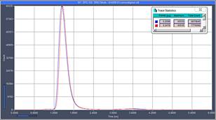
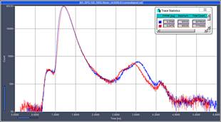
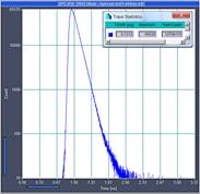
Fig. 1: Top: IRF of the two channels of the BiG-2 detector recorded by the
SPC-150 modules of the Simple-Tau system. Left: Linear scale. Right:
Logarithmic scale. Horizontal scale 0 to 6 ns. The IRF width is about
250 ps. Bottom: IRF of the HPM‑100-40 detector for comparison.
Horizontal scale 0 to 3.3 ns, diagrams are at same time scale as Fig. 1,
top. The IRF of the HPM is 130 ps.
The full-width at half maximum (FWHM) is
about 250 ps. The logarithmic display (top right) shows that there is a slight
shoulder before the main peak, and a secondary peak about 1.5 ns later. Such
secondary IRF structures are a common feature of conventional PMTs [1]. Their
presence in the BiG-2 detector is therefore not surprising. The amplitude of
the sub-structures is moderate and has little influence on the accuracy of
fluorescence lifetime analysis. It may, however, complicate the extraction of
with low-amplitude decay components from double- or triple-exponential decay profiles.
Fig. 1, bottom shows the IRF of an HPM‑100-40
hybrid detector at the same time scale. The HPM‑100-40 is the standard
detector for the bh FLIM systems, and still the gold standard in IRF shape,
time-resolution and efficiency [5]. A comparison of the curves shows that the
HPM has a narrower IRF (130 ps FWHM) and no secondary peaks.
NDD FLIM with two-Photon Excitation
NDD FLIM images were recorded with an
LSM710/780 OPO system [4]. Lifetime
images recorded from a covallaria sample are shown in Fig. 2, left. The
photons were detected in a wavelength interval from 575 to 610 nm. The
image is free of optical reflections or other artefacts. The decay data are
clean, without any traces of optical reflections or leakage of laser light, see
Fig. 2, right.
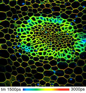
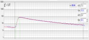
Fig. 2: Left: NDD FLIM image of a convallaria sample. Right: Decay data in
selected pixel of the image. Excitation by OPO at 1100 nm, detection at 575
to 610 nm. Data format 512x512 pixels, 256 time channels.
Amplitude-weighted lifetime of double-exponential decay, incomplete decay model
[1,2].
A more challenging test for any laser
scanning microscope is imaging of thick samples of high internal scattering. We
therefore stained a sample of fresh pig skin with methylen blue, and recorded
images with 2-photon excitation at 1100 nm. The BiG-2 detector passed this
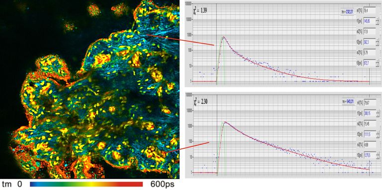
test without problems. The result is shown in Fig. 3. The lifetime image is
shown on the left, decay curves from two selected areas on the right.
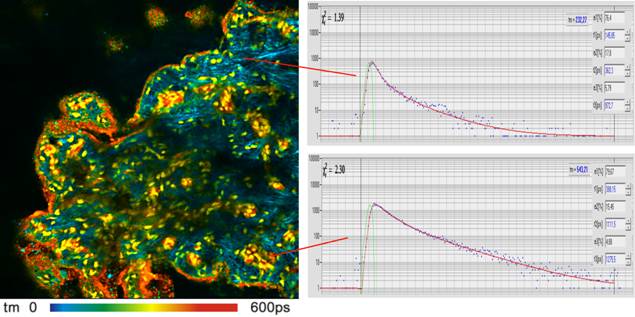
Fig. 3: Fresh pig skin stained with methylen blue. Left: FLIM image,
512x512 pixels, amplitude-weighted lifetime of triple-exponential decay. Right:
Fluorescence decay curves in two selected spots of the image. Two-photon
excitation by OPO at 1100 nm, non-descanned detection by BiG-2 detector.
Confocal FLIM with One-Photon Excitation
Confocal FLIM was tested in an LSM 880
with a 405 nm picosecond diode laser. In the LSM 880 the confocal
output from the scan head is used for the Airy-Scan detector. It is therefore
not directly accessible. For additional confocal detectors Zeiss offers a beam
switch that is inserted between the output of the scan head and the Airy Scan
detector, see Fig. 4, left. The 90° position of the switch directs the beam to
a vertical (BiG-type) port. This port can be used both for the BiG-2 detector and
for the bh hybrid detectors, see Fig. 4, right.
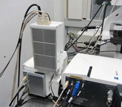
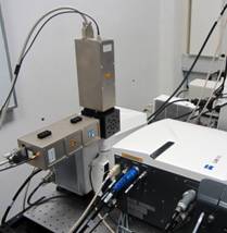
Fig. 4: LSM 880 scan head with BiG-2 detector (left) and with two bh
HPM‑100 hybrid detectors (right). The detectors are connected to a beam
switch between the Airy Scan detector and the scan head.
Fig. 5 shows FLIM images of a convallaria
sample excited at 405 nm. The images were detected by the two spectral channels
of the BiG-2 detector, and recorded by the two parallel TCSPC channels of the
Simple-Tau 152 FLIM system.
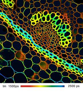
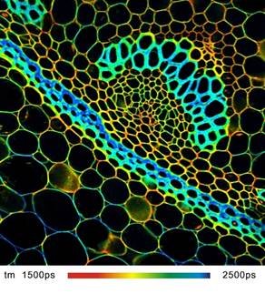
Fig. 5: Confocal FLIM images recorded by BiG-2 detector and LSM 880.
Left: Channel from 500 to 550 nm. Right: Channel from 575 nm to
610 nm. Convallaria sample, excitation by 405 nm ps diode laser,
512x512 pixels, 256 time channels. bh Simple-Tau 152 dual-channel TCSPC
FLIM system.
Images recorded in this setup were found
free of any artefacts. The sensitivity of the system was excellent. A
comparison of the BiG-2 with the HPM‑100-40 hybrid detectors (setup shown
in Fig. 4) showed no difference in efficiency.
FCS
FCS recording is a sensitive test for
detection efficiency. We therefore used the FLIM system to record FCS from a
number of dye solutions with the BiG-2 detector. The results were excellent. Fig.
6 shows decay curves and FCS curves of Atto 425 excited with the
405 nm ps diode laser. FCS with 405 nm excitation is not trivial.
Problems can especially occur by fluorescence of optical components, by leakage
of laser spectral background, and by poor transmission of the optics. Not so
with the LSM 880 - both decay curves and FCS curves were recorded at high
efficiency and without any noticeable artefacts.
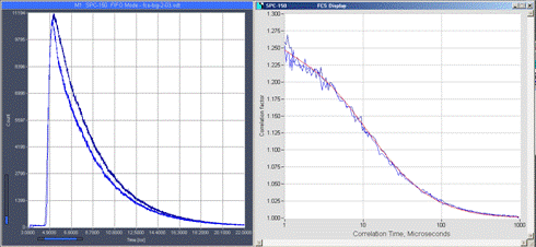
Fig. 6: Fluorescence decay functions and FCS curves detected in the two
channels of the BiG‑2 detector. Atto 425, excitation by 405 nm
ps diode laser. bh Simple-Tau 152 dual-channel FLIM system. The red curve
is a fit with two diffusion terms and a triplet term.
The bh TCSPC systems are able to record FCS
also CW lasers [1]. Fig. 7 shows an example. It shows an FCS curve of
Rhodamine 110, excited by the 488 nm CW laser of the LSM 880.
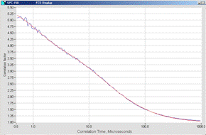
Fig. 7: FCS of Alexa 488 excited by 488 nm CW laser. Blue: FCS
data. Red: fit with two diffusion terms and a triplet term.
Conclusions
The BiG-2 detector in combination with the
bh FLIM systems delivers lifetime data with excellent sensitivity and
reasonably good time resolution. We did not find a noticeable sensitivity
difference to the bh HPM‑100 hybrid detectors [5]. Compared to the HPM detectors
the IRF is about a factor of 2 wider, and not entirely free of secondary peaks.
For fluorescence decay time longer than 400 ps this has little influence
on the lifetime accuracy. The secondary peak may, however complicate the
analysis of multi-exponential decay profiles with low-amplitude components.
Due to its excellent efficiency the BiG
detector works well also in FCS applications. The afterpulsing of the BiG-2 we
tested was surprisingly low. It was difficult to discern from the triplet
component of the FCS data.
In summary, it can be concluded that the BiG-2
is an excellent detector for steady-state imaging and a very useful detector
for FLIM. The advantage of the BiG-2 is that detectors need no be swapped when
a microscope port is used both for FLIM and for conventional imaging. However,
for high-end FLIM, such as double-exponential FRET measurements or metabolic
imaging via the fluorescence NADH the hybrid detectors still deliver superior
results. If such experiments have to be performed we recommend to keep a set of
hybrid detectors in reserve and attach them instead of the BiG-2 when needed.
The mechanical adapters of the hybrid PMTs and the BiG-2 are identical. The
detectors can thus be swapped by just loosening a single screw.
References
1. W. Becker, The bh TCSPC handbook. Becker & Hickl GmbH, 6th
Edition (2015), www.becker-hickl.com, printed copies available from bh
2. Becker & Hickl GmbH, Modular FLIM systems for Zeiss
LSM 510 and 710 family laser scanning microscopes. User handbook. www.becker-hickl.com, printed copies available from bh
3.
FLIM Systems for Zeiss LSM 710 Family Laser
Scanning Microscopes, an Overview. 40 pages, www.becker-hickl.com, printed
copies available from bh
4.
Multiphoton NDD FLIM at NIR Detection
Wavelengths with the Zeiss LSM 7MP and OPO Excitation. Application note,
www.becker-hickl.com
5. W. Becker, B. Su, K. Weisshart, O. Holub, FLIM and FCS Detection in
Laser-Scanning Microscopes: Increased Efficiency by GaAsP Hybrid Detectors.
Micr. Res. Tech. 74, 804-811 (2011)
Contact: Wolfgang Becker,
becker@becker-hickl.com