Principles
Perfusion Measurement by Diffuse Optical Correlation (DCS)
Perfusion measurements in tissue can be performed by correlating intensity fluctuations of photons scattered in the tissue A laser injects light into the tissue via a fibre. The light at a different spot is collected by another fibre and detected by a photon counting detector. An autocorrelation function of the intensity is calculated from the absolute times of the photons. To obtain a useful correlation function it is required that the laser have a coherence length longer than the average path length in the tissue, and that the fibres have diameters on the order of 20 µm or less. A simple DCS setup with a bh TCSPC module is shown in the figure below.
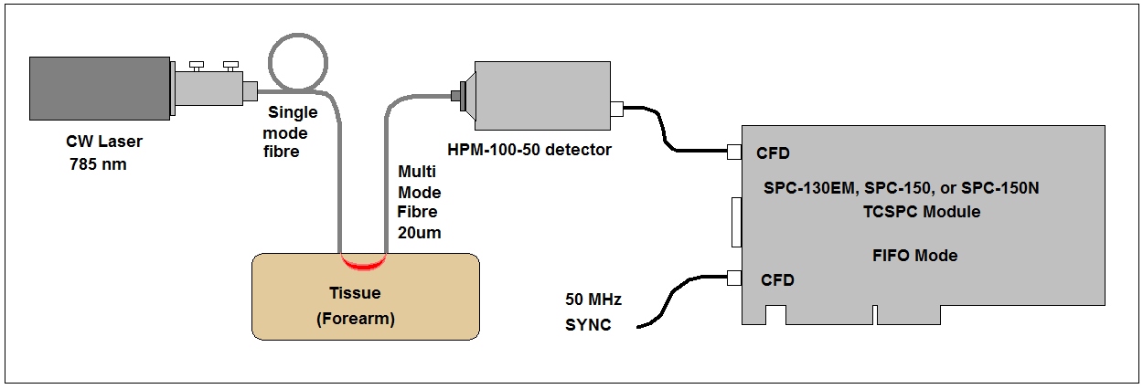
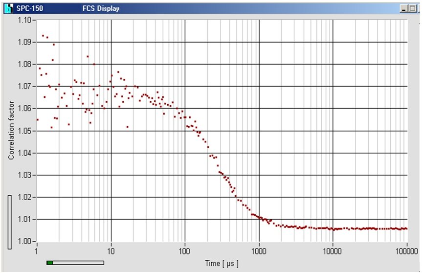
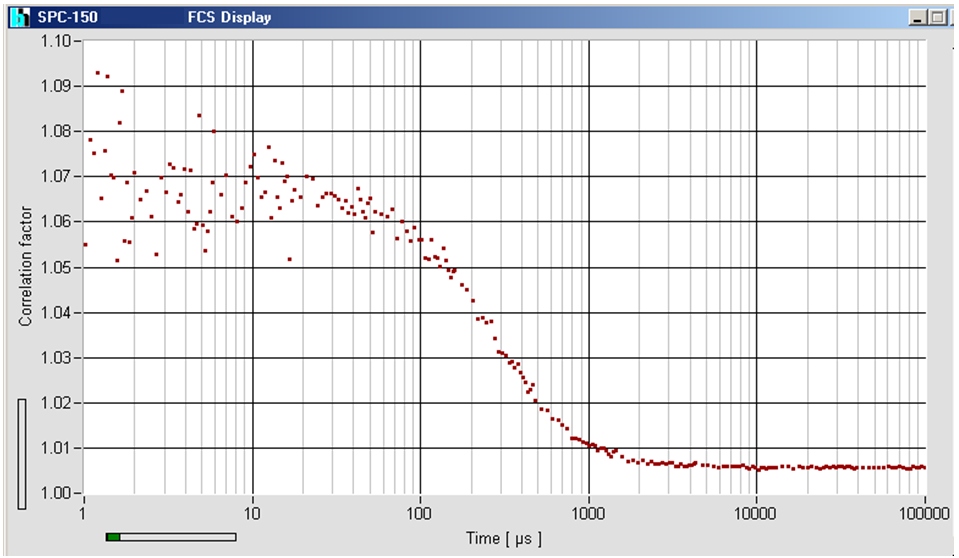
The laser is injected into the tissue by a single-mode fibre. Diffusely scattered light is collected by a 20 µm multi-mode fibre and detected by a HPM-100-50 hybrid detector. The photons are recorded by an SPC-130EM, SPC-150, or SPC-150N card in the FIFO mode. An autocorrelation function is calculated by the online correlation function of the SPCM software (see TCSPC Handbook). A result is shown in the figure below.
The setup can easily be extended to 8 detection channels by using a router in front of the SPC-150 card. DCS in 16 channels can be obtained by a bh DPC-230 correlator card. For more information, related applications and references, please see
The bh TCSPC Handbook, chapter ‘Diffuse Optical Tomography: DOT, NIRS and fNIRS’.