DCS-120 MP System Records Multiphoton FLIM
and PLIM
The DCS‑120 MP is an extended version
of the bh DCS‑120 confocal scanning FLIM System. It uses multiphoton excitation
by a femtosecond titanium-sapphire laser, fast galvanometer scanning,
non-descanned detection, hybrid detector technology, and single-photon
recording by bhs multi-dimensional TCSPC process. An AOM is included to
control the laser power and to modulate the laser for PLIM acquisition. The
system records FLIM data in two fully parallel recording channels, runs Z
stacks, accumulates fast FLIM time series, and records simultaneously FLIM and
PLIM. All components, including the laser and the AOM, are controlled by bhs
SPCM 64 bit data acquisition software. By using bhs 64 bit Megapixel FLIM technology,
images of the full field of view of the microscope can be recorded at
diffraction-limited resolution. Image formats as large as 2048 x 2048
pixels with 256 time channels per pixel are available.
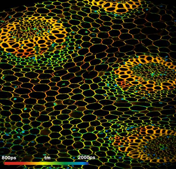
Convallaria sample with 1024 x 1024
pixels, 256 time channel per pixel. DCS‑120 scan
head, Nikon Eclipse inverted microscope, Spectra Physics Mai Tai laser. Microscope
lens 20x NA = 0.5. Excitation wavelength 800 nm
Due to its fast scan rates and its high
sensitivity, the DCS-120 MP is compatible with live cell and life tissue
imaging. Typical applications are measurements of local molecular environment
parameters, protein interaction experiments by FRET, imaging of metabolic
parameters derived from the fluorescence decay functions of endogenous fluorophores,
and correlated metabolic and oxygen saturation imaging.
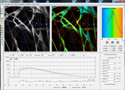
Phosphorescence
Lifetime Image, recorded by bhs PLIM technique based on laser modulation and
dual-time-base recording. SPCImage FLIM / PLIM data analysis.
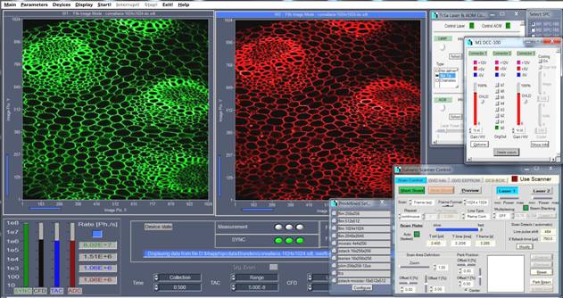
Main Panel of
SPCM Software. Images in two spectral channels, control panels for scanner,
laser and AOM, detectors, and predefined setup panel for easy selection of
imaging mode.
Key Specifications
Excitation Femtosecond
Titanium:Sapphire Laser
Wavelength Typ.
750 to 980 nm, depends on laser
Excitation
pulse frequency 75 to 80 MHz, depends on laser
Coupling into
scan head Free beam
Power control Acousto-optical
modulator (AOM). AOM is optional for FLIM but required for 2p PLIM. AOM can be
retrofitted.
Laser modulation
for PLIM Acousto-optical modulator (AOM).
AOM response
time 200 ns in PLIM mode
Laser and AOM
control via SPCM TCSPC/FLIM data acquisition
software
Additional excitation sources ps
diode lasers, supercontinuum laser with AOTF. Optional, can be retrofitted to
the system.
Microscopes All
inverted microscopes of Zeiss, Nikon, and Olympus
Detection
beam path Non-descanned (direct)
detection for 2p excitation
Optional
transmission path for SHG recording
2
confocal detection channels for 1p excitation
Piezo
actuators for confocal alignment
Detectors Two
HPM‑100-40 GaAsP hybrid detectors. No afterpulsing background, no
secondary pulses in IRF
Option:
HPM-100-50 GaAs hybrid detectors
Option:
MW-FLIM GaAsP 16-wavelength detector
Detector
protection Electronic overload
shutdown
Detection
wavelength selection Beamsplitter / filter cube in front of
detectors
Scanner bh
DCS-120 scan head
Alignment via
internal piezo actuators
Scanner
control integrated in SPCM
TCSPC/FLIM data acquisition software
Scan format,
pixels 2048 x 2048 1024
x 1024 512 x 512 256 x 256
Scan format,
time channels (max) 256 1024 4096 4096
Scan rate,
frames per sec., at zoom 4 0.37 0.65 1.47 2.95
Scan rate,
lines per sec., at zoom 4 500 750 750 750
Additional
scanner ports Additional port for visible-wavelength
laser
Two
outputs for additional confocal detectors
TCSPC System Two
parallel SPC-150, SPC-150N or SPC-160 channels
Upgrade
to three or four parallel channels possible
FLIM modes X-Y
scan, fast intensity preview mode, fast lifetime preview mode, Z Stack by record-and-save
procedure, Z Stack by Mosaic FLIM function, time series FLIM by record-and-save
procedure, time series FLIM by Mosaic FLIM function, fast accumulated Mosaic
FLIM time series, fast accumulated line scan time series (FLITS), PLIM,
simultaneous FLIM and PLIM
FCS mode Online
FCS, by correlating photon macro times, spot selected by beam park function of
scanner
Selection of operation mode Via
predefined-setup panel
FLIM data analysis By
bh SPCImage data analysis software. 1-2-3 exponential fit, incomplete-decay
model, 1st. moment analysis. No IRF recording necessary. Images of lifetime
components, amplitudes of components, intensity and amplitude-weighted
lifetime, relative intensity contribution, FRET efficiency. 1D histograms in
region of interest, 2D histograms of decay parameters, phasor plot.
For details,
please see Handbook of DCS-120 Confocal Scanning FLIM System, 6th ed. or later or
bh TCSPC Handbook, 6th ed. or later, both available for free download at
www.becker-hickl.com. Printed copies available from bh.
