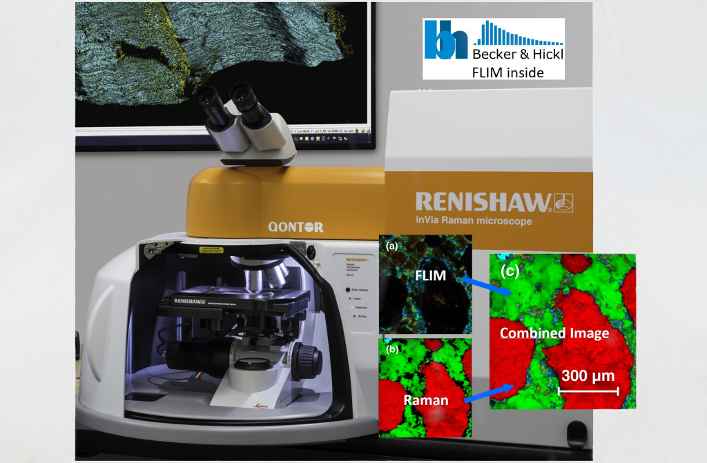
Renishaw’s renowned inViaTM Raman microscope is now available with Becker & Hickl’s top-of-the-class fluorescence lifetime imaging (FLIM) technique. This unique combination generates pixel-to-pixel correlated data, combining the power of FLIM and Raman.
FLIM and Raman Combined Images:
• Non-Invasive and Non-Destructive Raman and FLIM Imaging
• High Sensitivity and Efficiency with Renishaw’s Own Ultra-Low Noise, Ultra-High Sensitivity CCD Spectrometers and FLIM Detection at Single Photon Level
• High Resolution Confocal 2D and 3D Images
• Image Spatial Variation of Raman and FLIM Data
• Fully Automated
• Supports Multiple Lasers with Single-Click Rapid and Reliable Wavelength Change
• Maintain Focus in Real-Time with LiveTrack
• High-Speed Encoded Stage with Truly Confocal Spatial Resolution, Limited Only by the Inherent Diffraction Limit of Light
• Fast Fluorescence Imaging to Easily Identify and Define Locations for Subsequent Data Collection
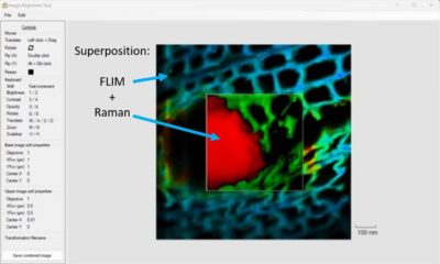
• Pixel-to-Pixel Correlation of FLIM and Raman Data
• Extend Raman Imaging to Sample Areas Dominated by Fluorescence
• Get Access to the Local Molecular Environment of the Fluorophore
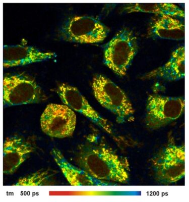
• Measure Cell Metabolic Health, Oxygen Concentration, PH, etc.
• Perform FLIM-FRET, FCS and Other Single Photon Counting Modalities
• Get Chemical and Structural Composition of the Sample
Description
FLIM and Raman Microscope
Renishaw’s renowned inViaTM Raman microscope is now available with Becker&Hickl’s top-of-the-class fluorescence lifetime imaging (FLIM) technique. This unique combination generates pixel-to-pixel correlated data, combining the power of FLIM and Raman. The obvious benefit for Raman practitioners is the ability to image sample regions dominated by fluorescence processes, in a much shorter time than a typical Raman image requires. Additionally, the BH FLIM addition goes far beyond conventional fluorescence intensity microscopy, providing access to the fluorescence lifetime in every pixel. This microscope enables studies matching the chemical and structural composition of a sample with information gained from FLIM data, e.g. metabolic cell health, oxygen concentration, local PH distribution, local temperature, the presence of certain marker proteins, etc.
Features
- Non-invasive and non-destructive imaging
- High sensitivity and efficiency
- Fully automated
- Supports multiple lasers with single-click rapid and reliable wavelength changing
- Maintain focus in real time with LiveTrack
- High-speed encoded stage
- Shared beam path for pixel-perfect correlation
- Pixel-to-pixel correlation of FLIM and Raman data performed in Renishaw WiRE™ software and Becker & Hickl SPC software
- Simple to set up and run correlative measurements
Raman Microscopy
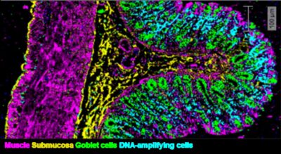
- Chemical and structural composition of the sample
- High sample specificity
Fluorescence Lifetime Imaging
- Access to sample areas dominated by fluorescence
- Fast imaging to find regions of interest
- Access to local molecular environment of the fluorophore
- Measure cell metabolic health, oxygen concentration, PH, etc.
- Perform FLIM-FRET, FCS and other single photon counting modalities