- For NSOM Systems of Nanonics and NT-MDT
- Recording by bh's Multidimensional TCSPC Technique
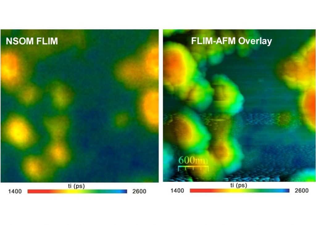
- Combines Atomic-Force and Fluorescence Lifetime Information
- High Sensitivity by HPM-100 Hybrid Detectors
- Excellent Time Resolution
- Excellent Timing Stability
- No Degradation of Time Resolution at Long Acquisition Times
- Fluorescence and Phosphorescence Lifetime Imaging
- FLIM Data Analysis by bh SPCImage NG
- Please See bh TCSPC Handbook, Section 'Time-Resolved Optical Near-Field Microscopy'
Description
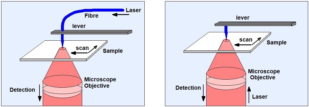
The scanning near-field optical microscope (SNOM or NSOM) combines the principles of the atomic force microscope and the laser scanning microscope. A sharp tip is scanned over the sample and kept in a distance comparable to the diameter of a single molecule. The tip can be the end of a tapered fibre through which the laser is fed to the sample, see figure below, left. Or, a metallic tip is illuminated by focusing the laser through the microscope objective on it. The evanescent field at the tip is used to probe the sample structure (Figure below, right). In both cases the fluorescence photons are collected through the microscope objective lens. Scanning is per-formed by a piezo-driven scan stage.
Because NSOM uses scanning it can easily be combined with bh TCSPC FLIM systems. The data are recorded by the usual imaging procedure of bh' SPCM data acquisition software. The data recording is synchronised with the scanner by the usual pixel, line, and frame clock pulses. No user-specific programming is necessary and the data can be processed by standard SPCImage NG data analysis software.
bh TCSPC FLIM systems are available for the NSOM systems of Nanonics and MD-MDT, please see corresponding application notes:
NSOM FLIM with the Nanonics AFM/NSOM System
An AFM/NSOM System with Fluorescence Lifetime Imaging
The FLIM systems can also be adapted to other NSOM systems, please see bh TCSPC Handbook.