- FLIM by bh's Multidimensional TCSPC technique
- Recording with STED-typical Resolution Down to 25 nm
- Recording of Standard bh FLIM Data Sets
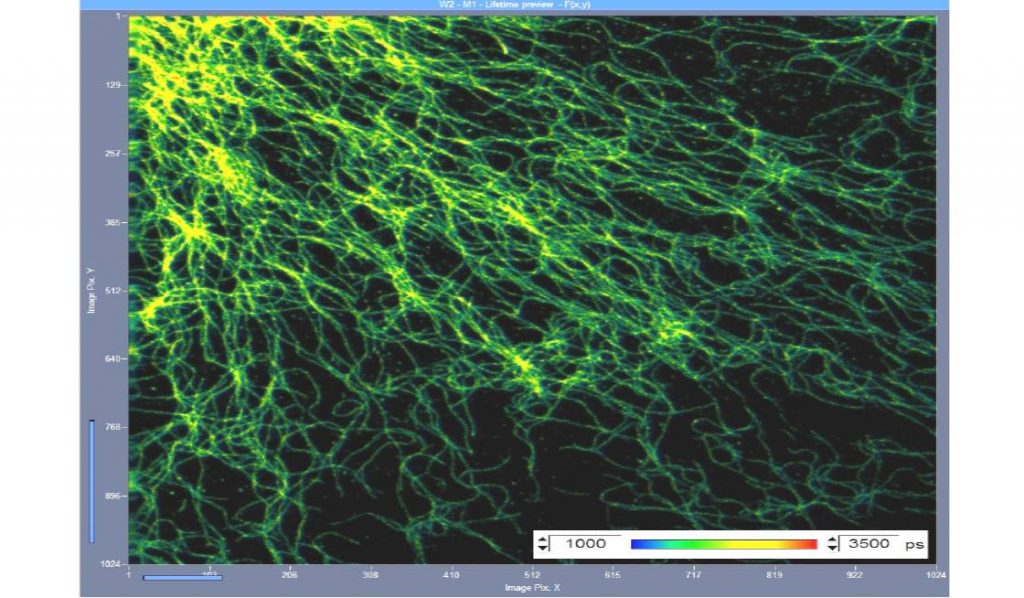
- Online-Lifetime Display by SPCM Data Acquisition Software
- Decay Analyis by SPCImage
- Time Gating to Suppress Undepleted Fluorescence
- Integrated in Abberior STED Microscopes
Description
Stimulated Emission Depletion (STED) microscopy exploits the nonlinearity of stimulated emission to obtain optical super-resolution. The fluorescence excited by the excita-tion beam in a scanning microscope is depleted by stimulated emission induced by a second (STED) laser beam. The wavefront of the STED beam is manipulated to obtain a diffraction pattern that either has doughnut shape laying in the x-y plane or a dumbbell shape oriented along the z axis. Fluorescence remains un-depleted in the centre part of the doughnut or the dumbbell. Because stimulated emission is highly nonlinear the un-depleted volume can be made considerably smaller than the point-spread function of the excitation beam. By scanning both beams together images with optical super-resolution are obtained. Because STED microscpy uses scanning it can be combined with TCSPC FLIM easily. All that is needed to combined the systems are the scan clock pulses, pixel, line and fram clock, from the scanner. Please see The bh TCSPC Handbook.
Downloads
Documents
Application Notes:
bh – Abberior Combination Records STED FLIM at Megapixel Resolution
Online STED FLIM with bh – Abberior Combination
Overview Brochures:
FLIM Systems for Laser Scanning Microscopes
The bh TCSPC Technique – Principles and Applications
Handbook: