bh FLIM Systems Record
Calcium Transients in Live Neurons
Abstract: We demonstrate the measurement of transient changes of
the Ca2+ concentration in live neurons by Fluorescence Transient
Lifetime Scanning (FLITS) and by temporal Mosaic FLIM. FLITS is based on the
build-up of a photon distribution over the distance along a line scan, the
times of the photons after the laser pulse, and the times of the photons after
a periodic stimulation of the sample, temporal mosaic FLIM on the buildup of a
photon distribution over the coordinates of a fast repetitive x-y scan, and the
photon times after the laser pulses and the stimulation pulses. For the
commonly used scanners the time resolution is about 1 ms for FLITS and
about 40 ms for temporal mosaic FLIM.
Ca2+ ions are involved in a
large number of cell functions, such as intracellular transport, membrane
potential, muscle contraction, gene expression, and cell differentiation. There
is a wide variety of Ca2+ sensors [4, 5, 6] which change their fluorescence lifetimes with the Ca2+
concentration in their local environment. Most likely, the mechanism of the Ca2+-dependent
lifetime change is that the fluorophore has a Ca-bound and a Ca-unbound form of
different fluorescence quantum efficiency and thus different fluorescence
lifetime. The fluorescence lifetime of the bound form is higher than that of
the unbound form. Consequently, the net fluorescence lifetime depends on the Ca2+
concentration. It can, however, happen that the fluorescence quantum efficiency
of the unbound form is so low that the corresponding lifetime component is no
longer observed. In that case, an intensity change but no lifetime change is
observed [5].
This is the case for the Fluo sensors, as has been shown for Fluo-4 [2]. However, the traditional Ca2+ dyes, such
as Calcium Green and Oregon Green, display large lifetime changes and work
beautifully for lifetime-based Ca2+ measurement. An example of a Ca2+
image is shown is Fig. 1.
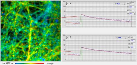
Fig. 1: FLIM image of cultured neurons stained with Oregon green OGB-1 AM.
Colour range from tm = 1200 ps (blue) to 2400 ps
(red). Decay curves of regions with low Ca (top) and high Ca (bottom) shown on
the right. Data courtesy of Inna Slutsky and Samuel Frere, Tel Aviv University,
Sackler School of Medicine.
The advantage of FLIM over intensity-based
Ca2+ imaging is that absolute values of the Ca2+
concentration are obtained. This has been used to quantify calcium
concentrations in astrocytes of live mice with cortical plaques by multiphoton NDD FLIM [3].
The Ca2+ concentration in cells
can change within remarkably short periods of time. Recording these Ca2+
transients requires a time resolution in the range of less than 50 ms. It
is thus usually considered impossible to record Ca2+ transients by
fluorescence lifetime detection. However, Ca2+ transients can easily
be recorded by using FLITS (fluorescence lifetime-transient scanning) or
temporal Mosaic FLIM [1].
FLITS builds up a photon distribution over
the distance within a line scan, the times of the photons in the fluorescence
decay, and the time after a stimulation of the sample. The application to the recording of
Ca2+ transients in live neurons on electrical stimulation has
been described in [2]. A typical result is shown in Fig. 2. Hippocamal cultures were prepared from newborn rats
and kept under physiological conditions for 12 to 18 days. The cultures were
then loaded with OGB-1 AM. A Zeiss LSM 7 MP multiphoton microscope with a
normal bh Simple-Tau 150 FLIM system was used to run the FLITS
experiments. The cells were stimulated periodically at a fraction of the line
clock frequency. To run the experiments, an intensity image was taken by the
LSM 7 MP, and an appropriate location for the line scan selected.
Then the LSM 7 MP was switched into the line scanning mode. The data
acquisition in the FLIM system, the scanning in the LSM 7 MP, and the
stimulation were started. The stimulation pulses of 1 ms duration were
applied to the cell culture in intervals of 3 seconds. Data acquisition was
continued over about 300 seconds, i.e. photons from about 100 stimulation
periods were accumulated. The result is shown in Fig. 2, left.
To verify that the FLITS experiment did not
cause cell damage or photobleaching a FLIM image was recorded after the FLITS experiment.
It is shown in Fig. 2, right. It does not show any cell damage or
photobleaching effects along the scanned line. It also shows that the Ca2+
concentration returned to the resting level, compare bottom of FLITS image on
the left.
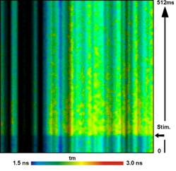
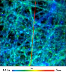
Fig. 2: FLITS of Ca2+
transients in live neurons. Left: FLITS image. Right: FLIM image taken after
the FLITS recording. Red lines indicates position of FLITS scan. Data courtesy of Inna Slutsky and Samuel Frere, Tel Aviv
University, Sackler School of Medicine.
A second
way to record fast temporal changes in the fluorescence behaviour of a sample
is temporal Mosaic FLIM. The technique records a photon distribution over the
coordinates of a fast repetitive x-y scan, the times of the photons after the
laser pulse, and the times of the photons after a stimulation of the sample [1]. The result is an extremely fast time series the
signal-to-noise ratio of which depends only on the total acquisition time but
not on the speed of the x-y scan. The technique became possible by bhs 64-bit
Megapixel technology which is able to record extremely large photon
distributions [1, 7].
Ca2+ recording by temporal
mosaic FLIM is shown in Fig.
3. OGB-1 AM was used as a Calcium sensor. The sample
was stimulated electrically every 3 seconds, and 100 stimulation cycles were accumulated.
A Zeiss LSM 7 MP was used for the experiment. With
64 x 64 pixels and a zoom factor of 5, the LSM 7 MP reaches a
frame time of 38 ms. 150 milliseconds before every stimulation a
recording through the entire 64-element mosaic was started. With the frame time
of 38 ms, the acquisition thus runs through the entire mosaic in 2.43
seconds. The result shows clearly the increase in the fluorescence lifetime of
the Ca2+ sensor in the mosaic elements 4 to 6, and a return to the
resting state over the next 10 to 15 mosaic elements (380 to 570 ms).
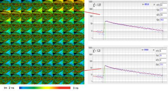
Fig. 3: Temporal mosaic FLIM of the Ca2+ transient in cultured
neurons after stimulation with an electrical signal. The time per mosaic
element is 38 milliseconds, the entire mosaic covers 2.43 seconds. Experiment
time runs from upper left to lower right. Photons were accumulated over 100
stimulation periods. Recorded by Zeiss LSM 7 MP and bh SPC‑150 TCSPC
module. Data courtesy of Inna Slutsky and Samuel Frere, Tel Aviv University,
Sackler Faculty of Medicine.
Conclusion
Ca2+ transients can be recorded
by using the FLITS or the Mosaic FLIM functions of the bh TCSPC FLIM systems.
The results are indendent of the spatially variable concentration of the Ca2+
sensor dye. Temporal changes in the Ca2+ concentration are recorded
at a resolution of about 1 ms by FLITS and about 40 ms by Mosaic
FLIM.
References
1.
W. Becker, The bh TCSPC
handbook. 6th edition. Becker & Hickl GmbH (2014), www.becker-hickl.com
2.
W. Becker, V. Shcheslavkiy, S.
Frere, I. Slutsky, Spatially Resolved Recording of Transient
Fluorescence-Lifetime Effects by Line-Scanning TCSPC. Microsc. Res. Techn. 77,
216-224 (2014)
3.
K.V.
Kuchibhotla, C.R. Lattarulo, B. Hyman, B. J. Bacskai, Synchronous hyperactivity
and intercellular calcium waves in astrocytes in Alzheimer mice. Science 323,
1211-1215
4.
Lakowicz
J.R., Szmacinski H., Johnson M.L., Calcium imaging using fluorescence lifetimes
an long-wavelength probes. J. Fluoresc. 2, 47-62 (1992)
5.
J.R. Lakowicz, Principles of
Fluorescence Spectroscopy, 3rd edn., Springer (2006)
6.
A. Minta, J.P.Y. Kao, R.Y.
Tsien, Fluorescent indicators for cytosolic calcium based on rhodamine and fluorescein
chromophores, J. Biol. Chem. 264, 8171-8178 (1989)
7.
H. Studier, W. Becker,
Megapixel FLIM. Proc. SPIE 8948 (2014)
