Principles
Ophthalmic FLIM (FLIO)
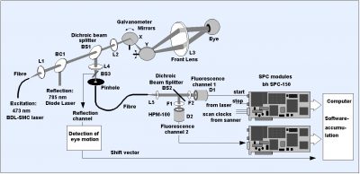
TCSPC FLIM systems are so sensitive that they are able to record lifetime images of the fundus (background) of the human eye. Ophthalmic FLIM systems use TCSPC FLIM in combination with fast beam scanning and excitation by a picosecond diode laser. The scanner of the Heidelberg Engineering FLIO system uses resonance scanning in x and galvanometer scanning in y. The scan rate is on the order of 16 frames per second. The laser beam is projected into the pupil of the eye through the front lens of the scan head. The fluorescence light returning from the eye background is descanned, projected into a pinhole, split into two wavelength channels, and detected by two HPM-100-40 GaAsP hybrid detectors. The photon pulses from the detectors are processed in two parallel SPC-150 TCSPC FLIM modules. In parallel with FLIM imaging, infrared reflection images are recorded in short intervals. These images are used to compensate eye motion in the FLIM recording. The principle is illustrated in the figure below.

Clinical Relevance
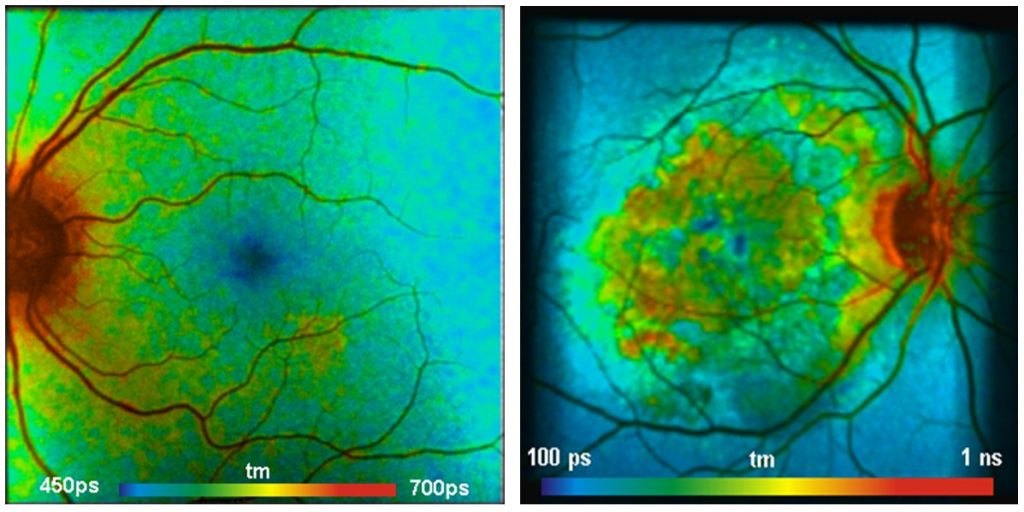
Early stages of eye diseases are often accompanied by metabolic changes in the fundus of the eye. These, in turn, cause changes in the fluorescence decay parameters of endogenous fluorophores. Ophthalmic FLIM is currently at the sage of clinical tests. The results show that the technique is able to show early stages of eye diseases before these are detectable by conventional methods, and, importantly, before they have caused permanent damage to the retina.

References
References for Ophthalmic FLIM
- Karl M. Andersen, Lydia Sauer, Rebekah H. Gensure, Martin Hammer, Paul S. Bernstein, Characterization of Retinitis Pigmentosa Using Fluorescence Lifetime Imaging Ophthalmoscopy (FLIO). TVST 7 No. 3 (2018)
- Dysli, G. Quellec, M. Abegg, M. N. Menke, U. Wolf-Schnurrbusch, J. Kowal, J. Blatz, O. La Schiazza, A. B. Leichtle, S. Wolf, M. S. Zinkernagel, Quantitative Analysis of Fluorescence Lifetime Measurements of the Macula Using the Fluorescence Lifetime Imaging Ophthalmoscope in Healthy Subjects. IOVS 55, 2107-2113 (2014)
- Dysli, M.Dysli, V. Enzmann, S. Wolf, M. S. Zinkernagel, Fluorescence Lifetime Imaging of the Ocular Fundus in Mice. IOVS 55, 7206-7215 (2014)
- Dysli, S. Wolf, K. Hatz, M. S. Zinkernagel, Fluorescence Lifetime Imaging in Stargardt Disease: Potential Marker for Disease Progression. Invest Ophthalmol Vis Sci. 57, 832-841, (2016)
- C. Dysli, S. Wolf, H.V. Tran, M.S. Zinkernagel, Autofluorescence lifetimes in patients with choroideremia identify photoreceptors in areas with retinal pigment epithelium atrophy. Invest Ophthalmol Vis Sci. 2016;57:6714–6721. DOI:10.1167/ iovs.16-20392
- Dysli, C., Wolf, S., Berezin, M.Y., Sauer, L., Hammer, M., Zinkernagel, M.S., Fluorescence lifetime imaging ophthalmoscopy, Progress in Retinal and Eye Research (2017), doi: 10.1016/j.preteyeres.2017.06.005
- J. B. Lincke, C Dysli, D Jaggi, R Fink, S. Wolf, M. S. Zinkernagel, The Influence of Cataract on Fluorescence Lifetime Imaging Ophthalmoscopy (FLIO). Transl Vis Sci Technol 10(4), 33, 1-10 (2021)
- Lydia Sauer, Rebekah H. Gensure, Karl M. Andersen, Lukas Kreilkamp, Gregory S. Hageman, Martin Hammer, Paul S. Bernstein, Patterns of Fundus Autofluorescence Lifetimes In Eyes of Individuals With Nonexudative Age-Related Macular Degeneration. IOVS 59 (2018)
- Lydia Sauer, Rebekah H. Gensure, PhD,1 Martin Hammer, Paul S. Bernstein, Fluorescence Lifetime Imaging Ophthalmoscopy: A Novel Way to Assess Macular Telangiectasia Type 2. Ophthalmology Retina 2 (6), 587-598 (2018)
- Lydia Sauer, Karl M. Andersen, Binxing Li, Rebekah H. Gensure, Martin Hammer, Paul S. Bernstein, Fluorescence Lifetime Imaging Ophthalmoscopy (FLIO) of Macular Pigment. Retina (2018)
- Lydia Sauer, Dietrich Schweitzer, Lisa Ramm, Regine Augsten, Martin Hammer, Sven Peters, Impact of Macular Pigment on Fundus Autofluorescence Lifetimes. Retina 56, 4668–4679 (2015)
- Schweitzer, S. Quick, S. Schenke, M. Klemm, S. Gehlert, M. Hammer, S. Jentsch, J. Fischer, Vergleich von Parametern der zeitaufgelösten Autofluoreszenz bei Gesunden und Patienten mit früher AMD. Der Ophthalmologe 8, 714-722 (2009)
- Schweitzer, Quantifying fundus autofluorescence. In: N. Lois, J.V. Forrester, eds., Fundus autofluorescence. Wolters Kluwer, Lippincott Willams & Wilkins (2009)
- Schweitzer, Metabolic Mapping. In: F.G. Holz, R.F. Spaide (eds), Medical retina, Essential in Opthalmology, Springer (2010)
- Schweitzer, S. Quck, M. Klemm, M. Hammer, S. Jentsch, J. Dawczynski, Zeitaufgelöste Autofluoreszenz bei retinalen Gefäßverschlüssen. Der Ophthalmologe 12, 1145-1152 (2010)
- Schweitzer, Autofluorescence diagnostics of ophthalmic diseases. In: V.V. Ghukasyan, A.H. Heikal, eds., Natural biomarkers for cellular metabolism. Biology, techniques, and applications. CRC Press, Taylor and Francis Group, Boca Raton, London, New York (2015)
- Schweitzer, M. Hammer, Fluorescence Lifetime Imaging in Ophthalmology. In: W. Becker (ed.) Advanced time-correlated single photon counting applications. Springer, Berlin, Heidelberg, New York (2015)
- Schweitzer, Ophthalmic applications of FLIM. In: L. Marcu. P.M.W. French, D.S. Elson, (eds.), Fluorecence lifetime spectroscopy and imaging. Principles and applications in biomedical diagnostics. CRC Press, Taylor & Francis Group, Boca Raton, London, New York (2015)
- Schweitzer, L. Deutsch, M. Klemm, S. Jentsch, M. Hammer, S. Peters, J. Haueisen, U. A. Müller, J. Dawczynski, Fluorescence lifetime imaging ophthalmoscopy in type 2 diabetic patients who have no signs of diabetic retinopathy. J. Biomed. Opt. 20(6), 061106-1 to 13 (2015)
