TCSPC Fibre-Probe System with an Exchangeable Tip
Wolfgang Becker, Ludwig Bergann, Becker & Hickl
GmbH, Berlin, Germany
Abstract: This application note describes a
fluorescence-lifetime detection system based on a fibre-optical probe with an exchangeable
tip. The excitation light is delivered to the tip via a single-mode fibre, the
emission light is transferred to the detector by a multi-mode fibre. The
electronic part of the system consist of a bh BDL-SMN picosecond diode laser, a
bh PMH‑100 hybrid detector or MW-FLIM GaAsP multi-wavelength detector,
and a Simple-Tau 150 TCSPC system. The system features high sensitivity
and short acquisition time. Clean fluorescence decay curves from a 10-7
mol/l fluorescein solution were recorded within an acquisition time of
0.5 seconds, time-series of autofluorescence decay curves were recorded at
a speed of 100 ms per step.
Fibre-optical probes in combination with
TCSPC have been described for a variety of tasks in spectroscopy of biological
tissue [1, 8, 9, 12]. Recently, Cui et al. [10] have implanted optical fibres
in the brains of mice to record behaviour-related Ca++ signals. A
limitation in these applications had been motion artefacts by variable speckle
patterns in the excitation fibres. Cui et al. solved this problem by using
single-mode fibres for excitation. The use of single-mode fibres, however,
leads to a problem with fibre-to-fibre coupling. The animals therefore could
not be freely connected and disconnected to or from the measurement system. A
solution was presented in [6, 11] in form of a novel miniature fibre-to-fibre
connection for single-mode fibres. A second way to solve the problem is to use
an implantable fibre tip that contains a short piece of multi-mode fibre [7].
It is connected to the single-mode source fibre and the multi-mode detection
fibre of the fibre-probe system by a single miniature fibre connector.
System Architecture
The instrument consists of the fibre probe,
the exchangeable tip, the excitation laser, the detector, and the TCSPC system.
The setup is shown in Fig. 1.
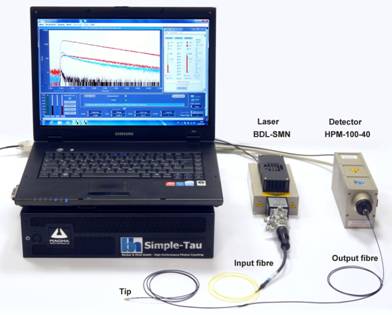
Fig. 1: Fibre-optical TCSPC system with fibre probe, BDL-SMN laser, HPM‑100-40
hybrid detector, and Simple-Tau 150 TCSPC system
The excitation light is delivered by a BDL‑SMN
picosecond diode laser [5]. It is injected into the input fibre of the fibre
probe. The fluorescence light returned from the measurement object is
transferred to an HPM‑100-40 detector [2, 3] by the output fibre of the probe. The
input fibre is single-mode to minimise motion artefacts. The output fibre is
multi-mode to obtain a high collection efficiency. The fluorescence decay
curves are recorded by a bh Simple‑Tau 150 TCSPC system [2].
The principle of the fibre probe [7] is
shown in Fig. 2. The probe consists of the input fibre (single mode) with a
Qioptiq compatible fibre connector, the output fibre (multi-mode) with an FC
connector, a miniature fibre connector, and the exchangeable tip. The tip contains
of a short piece of multi-mode fibre. The tip is the only part of the system
that is common for the excitation and the detection beam. Background signals
from fluorescence an Raman light generation in the glass of the probe are therefore
kept at an acceptably low level. Photos of the tip and the miniature fibre
connector are shown in Fig. 3.
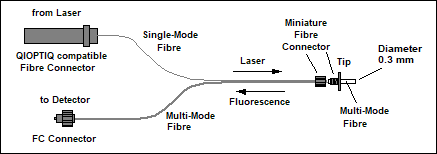
Fig. 2:
Principle of the fibre probe
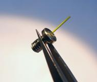
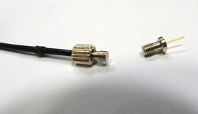
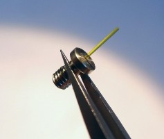
Fig. 3: Left:
Exchangeable tip. Right: Connection of the tip to the fibre system by miniature
fibre connector
Test Results
HPM‑100-40 Hybrid Detector
The system shown in Fig. 1 was tested with
fluorescein solutions of different concentration. For comparison, we also recorded
the autofluorescence signal from mammalian skin. Fig. 4, left and right, show
fluorescence decay curves of Fluorescein-Na 10-6 mol/l,
Fluorescein-Na 10-7 mol/l, autofluorescence of mammalian skin, and
the background fluorescence from the fibre probe. The excitation wavelength was
473 nm, the excitation power at the probe output was adjusted to
20 µW. The acquisition time was 0.5 seconds in Fig. 4, left, and 5
seconds in Fig. 4, right.
The results show that the probe background
is low enough to record clean data from a 10-7 mol/l
fluorescein solution. The signal obtained from the solution was approximately
at the level of the autofluorescence of mammalian skin. Both the
autofluorescence signal and the fluorescein signal were more than an order of
magnitude stronger than the background signal from the probe.
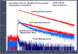
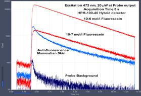
Fig. 4: Decay curves recorded with a HPM‑100‑40 hybrid
detector. Fluorescein-Na 10-6 mol/l, Fluorescein-Na 10-7
mol/l, autofluorescence of mammalian skin, background fluorescence from the
fibre probe. Excitation power 20 µW at probe output. Left: Acquisition
time 0.5 seconds. Right: Acquisition time 5 seconds.
It should be noted that these results were
obtained at an extremely low excitation power of 20 µW. This is a level
which is considered safe for in vivo measurements in biological systems. With
the BDL‑SMN diode laser the excitation power can, in principle, be
increased to more than 1 mW at the probe output.
MW FLIM GaAsP Multi-Wavelength Detector
The bh MW-FLIM family detectors (based on
PML‑16 16-channel detectors) record fluorescence decay curves in 16
wavelength intervals simultaneously [2, 4]. For multi-wavelength recording with
the fibre probe we used the MW FLIM GaAsP version with a Gallium-Arsenide-Phosphid
cathode. It has an efficiency about 5 time higher than the versions with
multi-alkali cathodes [2].
A 0.5-second recording from a 10-7
mol/l fluorescein solution is shown in Fig. 5, left, a recording of
autofluorescence from mammalian skin in Fig. 5, right. The excitation power was
20 µW at the tip output. The results show that even multi-spectral decay
data can be obtained at low excitation power and within a surprisingly short
acquisition time.
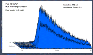
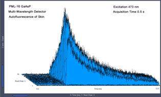
Fig. 5: Multi-wavelength decay data recorded with the MW FLIM GaAsP
detector. Left: Fluorescein-Na, 10-7 mol/l, acquisition time
0.5 seconds. Right: Autofluorescence of mammalian skin, acquisition time 0.5
seconds. Excitation 473 nm, 20 µW.
Time-Series Recording
The acquisition times achieved with the
system are short enough to record physiological changes in live objects at the
millisecond time scale. To demonstrate the feasibility of time-laps recording
we recorded a time-series of autofluorescence decays from human skin. Temporal
changes were induced by scanning the tip over the surface of the skin. The
excitation power was adjusted to 100 µW. Under these conditions, the TCSPC
system recorded about 106 photons per second. A recording rate of 10
measurements per second, i.e. a time per curve of 100 ms was used. A
typical result is shown in Fig. 6.
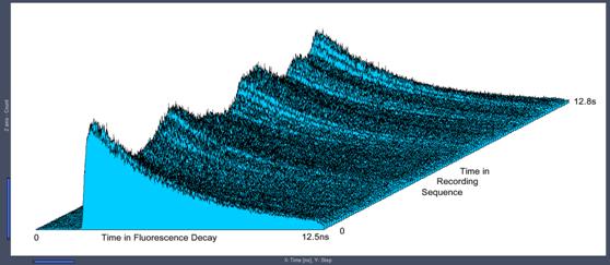
Fig. 6: Time-series
recorded at a rate of 100 ms per curve. Autofluorescence of skin, HPM‑100-40
hybrid detector, excitation power 100 µW.
The data shown in Fig. 6 contain about
100,000 photons per decay curve. The relative accuracy at which a fluorescence
lifetime can be derived from 100,000 photons is about 0.3 % [1, 2]. This
is much better than required for most bio-medical applications. For an accuracy
of 1%, only 10,000 photons per curve would be needed. That means, the speed of
the sequence can be increased to 100 curves per second without introducing
unacceptable variance in the recorded fluorescence lifetimes.
References
1.
W. Becker, Advanced time-correlated single-photon counting techniques. Springer (2005)
2.
W. Becker, The bh TCSPC handbook. 6th edition. Becker
& Hickl GmbH (2014), www.becker-hickl.com.
3.
Becker, W., Su, B., Weisshart, K. & Holub, O. (2011) FLIM and
FCS Detection in Laser-Scanning Microscopes: Increased Efficiency by GaAsP
Hybrid Detectors. Micr. Res. Tech. 74, 804-811
4.
Becker & Hickl GmbH, 16 channel
detector head for time-correlated single photon counting. User handbook,
www.becker‑hickl.com (2006)
5.
Becker & Hickl GmbH, BDL-SMN series
picosecond diode lasers. User handbook, www.becker-hickl.com
6.
Becker & Hickl GmbH, Implantable fibre-optical
fluorescence-lifetime detection system for in-vivo applications. Application
note, www.becker-hickl.com
7.
IFP-201 Implantable Fibre Probe for in vivo
Fluorescence Decay Measurements. Data sheet, www.becker-hickl.com
8.
P. A. A. De Beule, C. Dunsby, N. P. Galletly, G.
W. Stamp, A. C. Chu, U. Anand, P. Anand, C. D. Benham A. Naylor, P. M. W.
French, A hyperspectral fluorescence lifetime probe for skin cancer diagnosis.
Rev. Sci. Instrum. 78, 123101 (2007)
9.
S. Coda, A. J. Thompson,1,5 G. T. Kennedy, K. L.
Roche, L. Ayaru, D. S. Bansi, G. W. Stamp, A. V. Thillainayagam, P. M. W.
French, C. Dunsby, Fluorescence lifetime spectroscopy of tissue autofluorescence
in normal and diseased colon measured ex vivo using a fiber-optic probe.
Biomed. Opt. Expr. 5, 515-538 (2014)
10.
G. Cui, S.B.Jun, X. Jin, M.D. Pham, S.S. Vogel,
D.M. Lovinger, R.M. Costa, Concurrent activation of strial direct and indirect
pathways during action initiation. Nature 494, 238-242 (2013)
11.
G. Cui, S.B.Jun, X. Jin, G. Luo, M.D. Pham, D.M.
Lovinger, S.S. Vogel, R.M. Costa, Deep brain optical measurement of cell
type-specific neural activity in behaving mice. Nature Protocols, 9(6)
1213-1228 (2014)
12.
C. Dunsby, P.M.W. French, Single-point probes
for lifetime spectroscopy: Time-correlated single-photon counting technique.
In: L. Marcu. P.M.W. French, D.S. Elson, (eds.), Fluorecence lifetime
spectroscopy and imaging. Principles and applications in biomedical
diagnostics. CRC Press, Taylor & Francis Group (2015)